 Explore
 Validate
 Learn
Learn Western blot
Antibody data
- Antibody Data
- Antigen structure
- References [3]
- Comments [0]
- Validations
- Western blot [1]
- Immunoprecipitation [1]
- Immunohistochemistry [1]
Submit
Validation data
Reference
Comment
Report error
- Product number
- PAB10223 - Provider product page

- Provider
- Abnova Corporation
- Proper citation
- Abnova Corporation Cat#PAB10223, RRID:AB_1674726
- Product name
- EGFR polyclonal antibody
- Antibody type
- Polyclonal
- Description
- Rabbit polyclonal antibody raised against synthetic peptide of EGFR.
- Storage
- Store at 4°C. For long term storage store at -20°C.Aliquot to avoid repeated freezing and thawing.
Submitted references The EGF receptor provides an essential survival signal for SOS-dependent skin tumor development.
Role of epidermal growth factor receptor and STAT-3 activation in autonomous proliferation of SUM-102PT human breast cancer cells.
Occurrence of epidermal growth factor receptors in benign and malignant ovarian tumors and normal ovarian tissues: an immunohistochemical study.
Sibilia M, Fleischmann A, Behrens A, Stingl L, Carroll J, Watt FM, Schlessinger J, Wagner EF
Cell 2000 Jul 21;102(2):211-20
Cell 2000 Jul 21;102(2):211-20
Role of epidermal growth factor receptor and STAT-3 activation in autonomous proliferation of SUM-102PT human breast cancer cells.
Sartor CI, Dziubinski ML, Yu CL, Jove R, Ethier SP
Cancer research 1997 Mar 1;57(5):978-87
Cancer research 1997 Mar 1;57(5):978-87
Occurrence of epidermal growth factor receptors in benign and malignant ovarian tumors and normal ovarian tissues: an immunohistochemical study.
Henzen-Logmans SC, van der Burg ME, Foekens JA, Berns PM, Brussée R, Fieret JH, Klijn JG, Chadha S, Rodenburg CJ
Journal of cancer research and clinical oncology 1992;118(4):303-7
Journal of cancer research and clinical oncology 1992;118(4):303-7
No comments: Submit comment
Supportive validation
- Submitted by
- Abnova Corporation (provider)
- Main image

- Experimental details
- Western blot using EGFR polyclonal antibody (Cat # PAB10223) shows detection of a band at ~170 KDa corresponding to human EGFR present in unstimulated (Lane 1) and EGF (50 ng/mL for 15 min) stimulated (Lane 2) A-431 whole cell lysates (arrowhead).Approximately 30 ug of lysate was resolved on a 4-20% Tris-Glycine gel by SDS-PAGE and transferred onto nitrocellulose.After blocking, the membrane was probed with the primary antibody diluted to 1:1,000.Reaction occurred overnight at 4°C followed by washes and reaction with a 1:10,000 dilution of IRDye® 800 conjugated Gt-a-Rabbit IgG (H&L) MX10 for 45 min at room temperature (800 nm channel, green).Molecular weight estimation was made by comparison to prestained MW markers in lane M (700 nm channel, red).IRDye® 800 fluorescence image was captured using the Odyssey® Infrared Imaging System developed by LI-COR.IRDye is a trademark of LI-COR, Inc.

Supportive validation
- Submitted by
- Abnova Corporation (provider)
- Main image

- Experimental details
- Combined immunoprecipitation and immunoblot using EGFR polyclonal antibody (Cat # PAB10223).Lysates were prepared from GN4 rat liver epithelial cells both with (+) EGF treatment for 15' at 100 ng/mL and without (-) the addition of EGF.The combination of immunoprecipitation and immunoblotting was performed using the EGFR polyclonal antibody (Cat # PAB10223) for immunoprecipitation (10 uL) followed by immunoblot detection using an anti-phosphotyrosine antibody (Panel A).This was repeated in reverse order using a 1:2000 dilution of EGFR polyclonal antibody for immunoblot (Panel B).Visualization occurred using an ECL system.Film exposure was approximately 1'.
- Validation comment
- Immunoprecipitation

Supportive validation
- Submitted by
- Abnova Corporation (provider)
- Main image
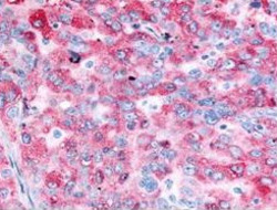
- Experimental details
- Immunohistochemical staining with EGFR polyclonal antibody (Cat # PAB10223) was diluted to 2.5 ug/mL detect EGFR in human ovarian cancer tissue. Tissue was formalin fixed and paraffin embedded. No pre-treatment of sample was required. The image shows the localization of antibody as the precipitated red signal, with a hematoxylin purple nuclear counter stain.
- Validation comment
- Immunohistochemistry (Formalin/PFA-fixed paraffin-embedded sections)
