 Explore
 Validate
 Learn
Learn Western blot
 Flow cytometry
Antibody data
- Antibody Data
- Antigen structure
- References [0]
- Comments [0]
- Validations
- Western blot [3]
- Immunohistochemistry [1]
Submit
Validation data
Reference
Comment
Report error
- Product number
- MA5-16360 - Provider product page

- Provider
- Invitrogen Antibodies
- Product name
- EGFR Monoclonal Antibody (SP84)
- Antibody type
- Monoclonal
- Antigen
- Synthetic peptide
- Description
- Heat-mediated antigen retrieval is recommended prior to staining, using a 10mM citrate buffer, pH 6.0, for 10 minutes followed by cooling at room temperature for 20 min. Following antigen retrieval, incubate samples with primary antibody for 30 min at room temperature. A suggested positive control is lung carcinoma or placenta tissue.
- Reactivity
- Human
- Host
- Rabbit
- Isotype
- IgG
- Antibody clone number
- SP84
- Vial size
- 500 µL
- Storage
- Store at 4°C short term. For long term storage, store at -20°C, avoiding freeze/thaw cycles.
No comments: Submit comment
Supportive validation
- Submitted by
- Invitrogen Antibodies (provider)
- Main image
- Experimental details
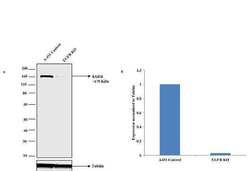
- Western blot analysis of EGFR (Fig a) was performed by loading 30 µg of A-431 Control (lane1), A-431 EGFR knockout (lane 2) whole cell extracts using Novex® NuPAGE® 10 % Bis-Tris gel, XCell SureLock™ Electrophoresis System (EI0002), Novex® Sharp Pre-Stained Protein Standard (LC5800), and iBlot® Dry Blotting System (IB21001). Proteins were transferred to a nitrocellulose membrane and blocked with 5% skim milk for 1 hour at room temperature. EGFR was detected at ~ 170 kDa using Rabbit anti-EGFR Monoclonal Antibody (Product # MA5-16360) at 1:1000 in 5% skim milk at 4°C overnight on a rocking platform. Goat anti-Rabbit IgG (H+L) Superclonal™ Secondary Antibody HRP conjugate (Product # A27036, 1:4000 dilution) was used and chemiluminescent detection was performed using Pierce™ ECL Western Blotting Substrate (Product # 32106). Densitometric analysis of this western blot is shown in histogram (Fig b). Reduction of signal in CRISPR mediated knockout (KO) confirms that antibody is specific to EGFR.

- Submitted by
- Invitrogen Antibodies (provider)
- Main image

- Experimental details
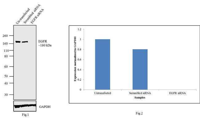
- Knockdown of EGFR was achieved by transfecting A549 cells with EGFR specific validated siRNAs (Silencer® select Product # s563, s564, s565). Western blot analysis (Fig. 1) was performed using membrane enriched extracts from the EGFR knockdown cells (lane 3), non-specific scrambled siRNA transfected cells (lane 2) and untransfected cells (lane 1). The blots were probed with Anti-EGFR Antibody, Rabbit Monoclonal (Product # MA5-16360, 1 µg/mL) and Goat anti-Rabbit IgG (H+L) Superclonal™ Secondary Antibody, HRP conjugate (Product # A27036, 0.25 µg/mL, 1:4000 dilution). Densitometric analysis of this western blot is shown in histogram (Fig. 2). Decrease in signal upon siRNA mediated knock down confirms that antibody is specific to EGFR.
- Submitted by
- Invitrogen Antibodies (provider)
- Main image

- Experimental details
- Western blot analysis was performed on membrane enriched extracts (30 µg lysate) of A-431 (Lane 1), H1975 (Lane 2), A549 (Lane 3), HeLa (Lane 4), U-87 MG (Lane 5) and MDA-MB-231 (Lane 6). The blots were probed with Anti-EGFR Monoclonal Antibody (Product # MA5-16360, 1:25 dilution) and detected by chemiluminescence using Goat anti-Mouse IgG (H+L) Superclonal™ Secondary Antibody, HRP conjugate (Product # A28177, 0.25 µg/mL, 1:4000 dilution). A 170 kDa band corresponding to EGFR was observed across the cell lines tested. Known quantity of protein samples were electrophoresed using Novex® NuPAGE® 4-12 % Bis-Tris gel (Product # NP0321BOX), XCell SureLock™ Electrophoresis System (Product # EI0002) and Novex® Sharp Pre-Stained Protein Standard (Product # LC5800). Resolved proteins were then transferred onto a nitrocellulose membrane using the wet transfer system. The membrane was probed with the relevant primary and secondary antibody following blocking with 5 % skimmed milk. Chemiluminescent detection was performed using Pierce™ ECL Western Blotting Substrate (Product # 32106).

Supportive validation
- Submitted by
- Invitrogen Antibodies (provider)
- Main image
- Experimental details
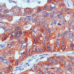
- Immunohistochemical analysis of Epidermal Growth Factor Receptor using anti-Epidermal Growth Factor Receptor Monoclonal Antibody (Product # MA5-16360) in Lung Squamous cell carcinoma Cancer Tissue. The recommened dilution for this antibody in immunohistochemistry applications is 1:100.
