 Explore
 Validate
 Learn
LearnMA5-13269
antibody from Invitrogen Antibodies
Targeting: EGFR
ERBB, ERBB1
 Western blot Immunocytochemistry
 Immunoprecipitation Immunohistochemistry Flow cytometry Other assay
Antibody data
- Antibody Data
- Antigen structure
- References [71]
- Comments [0]
- Validations
- Western blot [4]
- Immunocytochemistry [3]
- Immunohistochemistry [1]
- Flow cytometry [1]
- Other assay [4]
Submit
Validation data
Reference
Comment
Report error
- Product number
- MA5-13269 - Provider product page

- Provider
- Invitrogen Antibodies
- Product name
- EGFR Monoclonal Antibody (111.6)
- Antibody type
- Monoclonal
- Antigen
- Recombinant full-length protein
- Description
- MA5-13269 targets Epidermal Growth Factor Receptor in IHC (P), IP, and WB applications and shows reactivity with Human samples. The MA5-13269 immunogen is extracellular domain of human recombinant EGFR protein.
- Reactivity
- Human, Mouse, Rat
- Host
- Mouse
- Isotype
- IgG
- Antibody clone number
- 111.6
- Vial size
- 500 µL
- Concentration
- 0.2 mg/mL
- Storage
- 4° C
Submitted references Golgi Acidification by NHE7 Regulates Cytosolic pH Homeostasis in Pancreatic Cancer Cells.
β-Catenin/CBP inhibition alters epidermal growth factor receptor fucosylation status in oral squamous cell carcinoma.
Exosomes derived from oviduct cells mediate the EGFR/MAPK signaling pathway in cumulus cells.
A Chlamydia pneumoniae adhesin induces phosphatidylserine exposure on host cells.
Interaction of the EGFR signaling pathway with porcine cumulus oocyte complexes and oviduct cells in a coculture system.
Multiplexed Molecular Imaging of Biomarker-Targeted SERS Nanoparticles on Fresh Tissue Specimens with Channel-Compressed Spectrometry.
Quantitative molecular phenotyping with topically applied SERS nanoparticles for intraoperative guidance of breast cancer lumpectomy.
Missense Mutations in Exons 18-24 of EGFR in Hepatocellular Carcinoma Tissues.
An early endosome regulator, Rab5b, is an LRRK2 kinase substrate.
Quantification of the binding potential of cell-surface receptors in fresh excised specimens via dual-probe modeling of SERS nanoparticles.
EGFR, HER-2 and KRAS in canine gastric epithelial tumors: a potential human model?
EGFR, HER2 and HER3 dimerization patterns guide targeted inhibition in two histotypes of esophageal cancer.
Role of EGFR as a prognostic factor for survival in head and neck cancer: a meta-analysis.
Integrins and their ligands are expressed in non-small cell lung cancer but not correlated with parameters of disease progression.
Single cell stochastic regulation of pilus phase variation by an attenuation-like mechanism.
No-cost manual method for preparation of tissue microarrays having high quality comparable to semiautomated methods.
Receptor tyrosine kinase inhibitor profiling using bead-based multiplex sandwich immunoassays.
Identification of different subtypes of breast cancer using tissue microarray.
Epidermal growth factor receptor as a biomarker for cervical cancer.
Transdifferentiation of glioblastoma cells into vascular endothelial cells.
An aCGH classifier derived from BRCA1-mutated breast cancer and benefit of high-dose platinum-based chemotherapy in HER2-negative breast cancer patients.
Expression of epidermal growth factor receptor and vascular endothelial growth factor in vaginal squamous cell cancer.
MAP Kinase activation by receptor tyrosine kinases: in control of cell migration.
Triple-negative breast cancer: immunohistochemical correlation with basaloid markers and prognostic value of survivin.
Comparison of four antibodies for immunohistochemical evaluation of epidermal growth factor receptor expression in non-small cell lung cancer.
Prostaglandin E1 and misoprostol increase epidermal growth factor production in 3D-cultured human annulus cells.
Structure-function analysis of nucleolin and ErbB receptors interactions.
EGFR and ADAMs cooperate to regulate shedding and endocytic trafficking of the desmosomal cadherin desmoglein 2.
Ultraviolet irradiation-induces epidermal growth factor receptor (EGFR) nuclear translocation in human keratinocytes.
TGFalpha and EGFR in ovine preimplantation embryos and effects on development.
The role of heparin-binding EGF-like growth factor and amphiregulin in the epidermal proliferation of psoriasis in cooperation with TNFalpha.
Engineering of PDMS surfaces for use in microsystems for capture and isolation of complex and biomedically important proteins: epidermal growth factor receptor as a model system.
Sex steroid and epidermal growth factor profile of giant meningiomas associated with pregnancy.
Gene expression profiling and histopathological characterization of triple-negative/basal-like breast carcinomas.
Basaloid adenocarcinoma. A new variant of pulmonary adenocarcinoma.
Epidermal growth factor ligand/receptor loop and downstream signaling activation pattern in completely resected nonsmall cell lung cancer.
Dual-mode reflectance and fluorescence near-video-rate confocal microscope for architectural, morphological and molecular imaging of tissue.
Co-expression of EGF receptor, TGFalpha and S6 kinase is significantly associated with colorectal carcinomas with distant metastases at diagnosis.
Does the EGFR and VEGF expression predict the prognosis in colon cancer?
Molecular subtypes of breast cancer and amplification of topoisomerase II alpha: predictive role in dose intensive adjuvant chemotherapy.
Real-time detection of epidermal growth factor receptor expression in fresh oral cavity biopsies using a molecular-specific contrast agent.
Downregulation of EGF receptor signaling in pancreatic islets causes diabetes due to impaired postnatal beta-cell growth.
Sulindac metabolites inhibit epidermal growth factor receptor activation and expression.
Gene expression and immunolocalization of heparin-binding epidermal growth factor-like growth factor and human epidermal growth factor receptors in human corpus luteum.
Using liquid crystals to report membrane proteins captured by affinity microcontact printing from cell lysates and membrane extracts.
Epidermal growth factor receptor, protein kinase B/Akt, and glioma response to erlotinib.
Fluorescent nanocrystals for use in early cervical cancer detection.
Cyclooxygenase-2 (COX-2) levels before and after chemotherapy: a study in rectal cancer.
Cyclooxygenase-2 (COX-2) levels before and after chemotherapy: a study in rectal cancer.
A far-red fluorescent contrast agent to image epidermal growth factor receptor expression.
In vitro chemoresistance and biomarker profiles are unique for histologic subtypes of epithelial ovarian cancer.
In vitro chemoresistance and biomarker profiles are unique for histologic subtypes of epithelial ovarian cancer.
Activity of a specific inhibitor, gefitinib (Iressa, ZD1839), of epidermal growth factor receptor in refractory non-small-cell lung cancer.
Activity of a specific inhibitor, gefitinib (Iressa, ZD1839), of epidermal growth factor receptor in refractory non-small-cell lung cancer.
Inability of immunohistochemistry to predict clinical outcomes of endometrial cancer patients.
Inability of immunohistochemistry to predict clinical outcomes of endometrial cancer patients.
Characterization of a transplantable hormone-responsive human prostatic cancer xenograft TEN12 and its androgen-resistant sublines.
Geldanamycin-associated inhibition of intracellular trafficking is attributed to a co-purified activity.
Racial disparity of epidermal growth factor receptor expression in prostate cancer.
Domain-level antibody epitope mapping through yeast surface display of epidermal growth factor receptor fragments.
Validation of tissue array technology in head and neck squamous cell carcinoma.
Broadly distributed chemical reactivity of natural antibodies expressed in coordination with specific antigen binding activity.
A pharmacodynamic study of the epidermal growth factor receptor tyrosine kinase inhibitor ZD1839 in metastatic colorectal cancer patients.
Proliferation-differentiation relationships in the expression of heparin-binding epidermal growth factor-related factors and erbB receptors by normal and psoriatic human keratinocytes.
Optical systems for in vivo molecular imaging of cancer.
Optical systems for in vivo molecular imaging of cancer.
Profiling receptor tyrosine kinase activation by using Ab microarrays.
Retroperitoneal cystic papillary carcinoma of probable nephrogenic origin.
Retroperitoneal cystic papillary carcinoma of probable nephrogenic origin.
A sandwich type acridinium-linked immunosorbent assay (ALISA) detects soluble ErbB1 (sErbB1) in normal human sera.
A sandwich type acridinium-linked immunosorbent assay (ALISA) detects soluble ErbB1 (sErbB1) in normal human sera.
Galenkamp KMO, Sosicka P, Jung M, Recouvreux MV, Zhang Y, Moldenhauer MR, Brandi G, Freeze HH, Commisso C
Cancer discovery 2020 Jun;10(6):822-835
Cancer discovery 2020 Jun;10(6):822-835
β-Catenin/CBP inhibition alters epidermal growth factor receptor fucosylation status in oral squamous cell carcinoma.
Chandler KB, Alamoud KA, Stahl VL, Nguyen BC, Kartha VK, Bais MV, Nomoto K, Owa T, Monti S, Kukuruzinska MA, Costello CE
Molecular omics 2020 Jun 1;16(3):195-209
Molecular omics 2020 Jun 1;16(3):195-209
Exosomes derived from oviduct cells mediate the EGFR/MAPK signaling pathway in cumulus cells.
Lee SH, Oh HJ, Kim MJ, Lee BC
Journal of cellular physiology 2020 Feb;235(2):1386-1404
Journal of cellular physiology 2020 Feb;235(2):1386-1404
A Chlamydia pneumoniae adhesin induces phosphatidylserine exposure on host cells.
Galle JN, Fechtner T, Eierhoff T, Römer W, Hegemann JH
Nature communications 2019 Oct 11;10(1):4644
Nature communications 2019 Oct 11;10(1):4644
Interaction of the EGFR signaling pathway with porcine cumulus oocyte complexes and oviduct cells in a coculture system.
Lee SH, Oh HJ, Kim MJ, Setyawan EMN, Lee BC
Journal of cellular physiology 2019 Apr;234(4):4030-4043
Journal of cellular physiology 2019 Apr;234(4):4030-4043
Multiplexed Molecular Imaging of Biomarker-Targeted SERS Nanoparticles on Fresh Tissue Specimens with Channel-Compressed Spectrometry.
Kang S, Wang Y, Reder NP, Liu JT
PloS one 2016;11(9):e0163473
PloS one 2016;11(9):e0163473
Quantitative molecular phenotyping with topically applied SERS nanoparticles for intraoperative guidance of breast cancer lumpectomy.
Wang Y, Kang S, Khan A, Ruttner G, Leigh SY, Murray M, Abeytunge S, Peterson G, Rajadhyaksha M, Dintzis S, Javid S, Liu JT
Scientific reports 2016 Feb 16;6:21242
Scientific reports 2016 Feb 16;6:21242
Missense Mutations in Exons 18-24 of EGFR in Hepatocellular Carcinoma Tissues.
Panvichian R, Tantiwetrueangdet A, Sornmayura P, Leelaudomlipi S
BioMed research international 2015;2015:171845
BioMed research international 2015;2015:171845
An early endosome regulator, Rab5b, is an LRRK2 kinase substrate.
Yun HJ, Kim H, Ga I, Oh H, Ho DH, Kim J, Seo H, Son I, Seol W
Journal of biochemistry 2015 Jun;157(6):485-95
Journal of biochemistry 2015 Jun;157(6):485-95
Quantification of the binding potential of cell-surface receptors in fresh excised specimens via dual-probe modeling of SERS nanoparticles.
Sinha L, Wang Y, Yang C, Khan A, Brankov JG, Liu JT, Tichauer KM
Scientific reports 2015 Feb 26;5:8582
Scientific reports 2015 Feb 26;5:8582
EGFR, HER-2 and KRAS in canine gastric epithelial tumors: a potential human model?
Terragni R, Casadei Gardini A, Sabattini S, Bettini G, Amadori D, Talamonti C, Vignoli M, Capelli L, Saunders JH, Ricci M, Ulivi P
PloS one 2014;9(1):e85388
PloS one 2014;9(1):e85388
EGFR, HER2 and HER3 dimerization patterns guide targeted inhibition in two histotypes of esophageal cancer.
Fichter CD, Timme S, Braun JA, Gudernatsch V, Schöpflin A, Bogatyreva L, Geddert H, Faller G, Klimstra D, Tang L, Hauschke D, Werner M, Lassmann S
International journal of cancer 2014 Oct 1;135(7):1517-30
International journal of cancer 2014 Oct 1;135(7):1517-30
Role of EGFR as a prognostic factor for survival in head and neck cancer: a meta-analysis.
Keren S, Shoude Z, Lu Z, Beibei Y
Tumour biology : the journal of the International Society for Oncodevelopmental Biology and Medicine 2014 Mar;35(3):2285-95
Tumour biology : the journal of the International Society for Oncodevelopmental Biology and Medicine 2014 Mar;35(3):2285-95
Integrins and their ligands are expressed in non-small cell lung cancer but not correlated with parameters of disease progression.
Böger C, Kalthoff H, Goodman SL, Behrens HM, Röcken C
Virchows Archiv : an international journal of pathology 2014 Jan;464(1):69-78
Virchows Archiv : an international journal of pathology 2014 Jan;464(1):69-78
Single cell stochastic regulation of pilus phase variation by an attenuation-like mechanism.
Danne C, Dubrac S, Trieu-Cuot P, Dramsi S
PLoS pathogens 2014 Jan;10(1):e1003860
PLoS pathogens 2014 Jan;10(1):e1003860
No-cost manual method for preparation of tissue microarrays having high quality comparable to semiautomated methods.
Foda AA
Applied immunohistochemistry & molecular morphology : AIMM 2013 May;21(3):271-4
Applied immunohistochemistry & molecular morphology : AIMM 2013 May;21(3):271-4
Receptor tyrosine kinase inhibitor profiling using bead-based multiplex sandwich immunoassays.
Pötz O, Schneiderhan-Marra N, Henzler T, Herget T, Joos TO
Methods in molecular biology (Clifton, N.J.) 2012;795:191-202
Methods in molecular biology (Clifton, N.J.) 2012;795:191-202
Identification of different subtypes of breast cancer using tissue microarray.
Munirah MA, Siti-Aishah MA, Reena MZ, Sharifah NA, Rohaizak M, Norlia A, Rafie MK, Asmiati A, Hisham A, Fuad I, Shahrun NS, Das S
Romanian journal of morphology and embryology = Revue roumaine de morphologie et embryologie 2011;52(2):669-77
Romanian journal of morphology and embryology = Revue roumaine de morphologie et embryologie 2011;52(2):669-77
Epidermal growth factor receptor as a biomarker for cervical cancer.
Soonthornthum T, Arias-Pulido H, Joste N, Lomo L, Muller C, Rutledge T, Verschraegen C
Annals of oncology : official journal of the European Society for Medical Oncology 2011 Oct;22(10):2166-78
Annals of oncology : official journal of the European Society for Medical Oncology 2011 Oct;22(10):2166-78
Transdifferentiation of glioblastoma cells into vascular endothelial cells.
Soda Y, Marumoto T, Friedmann-Morvinski D, Soda M, Liu F, Michiue H, Pastorino S, Yang M, Hoffman RM, Kesari S, Verma IM
Proceedings of the National Academy of Sciences of the United States of America 2011 Mar 15;108(11):4274-80
Proceedings of the National Academy of Sciences of the United States of America 2011 Mar 15;108(11):4274-80
An aCGH classifier derived from BRCA1-mutated breast cancer and benefit of high-dose platinum-based chemotherapy in HER2-negative breast cancer patients.
Vollebergh MA, Lips EH, Nederlof PM, Wessels LFA, Schmidt MK, van Beers EH, Cornelissen S, Holtkamp M, Froklage FE, de Vries EGE, Schrama JG, Wesseling J, van de Vijver MJ, van Tinteren H, de Bruin M, Hauptmann M, Rodenhuis S, Linn SC
Annals of oncology : official journal of the European Society for Medical Oncology 2011 Jul;22(7):1561-1570
Annals of oncology : official journal of the European Society for Medical Oncology 2011 Jul;22(7):1561-1570
Expression of epidermal growth factor receptor and vascular endothelial growth factor in vaginal squamous cell cancer.
Brunner A, Grimm C, Polterauer S, Hefler L, Stani J, Dudek G, Horvat R
American journal of obstetrics and gynecology 2011 Feb;204(2):171.e1-6
American journal of obstetrics and gynecology 2011 Feb;204(2):171.e1-6
MAP Kinase activation by receptor tyrosine kinases: in control of cell migration.
Tarcic G, Yarden Y
Methods in molecular biology (Clifton, N.J.) 2010;661:125-35
Methods in molecular biology (Clifton, N.J.) 2010;661:125-35
Triple-negative breast cancer: immunohistochemical correlation with basaloid markers and prognostic value of survivin.
Dogu GG, Ozkan M, Ozturk F, Dikilitas M, Er O, Ozturk A
Medical oncology (Northwood, London, England) 2010 Mar;27(1):34-9
Medical oncology (Northwood, London, England) 2010 Mar;27(1):34-9
Comparison of four antibodies for immunohistochemical evaluation of epidermal growth factor receptor expression in non-small cell lung cancer.
Mathieu A, Weynand B, Verbeken E, Da Silva S, Decaestecker C, Salmon I, Demetter P
Lung cancer (Amsterdam, Netherlands) 2010 Jul;69(1):46-50
Lung cancer (Amsterdam, Netherlands) 2010 Jul;69(1):46-50
Prostaglandin E1 and misoprostol increase epidermal growth factor production in 3D-cultured human annulus cells.
Gruber HE, Hoelscher G, Loeffler B, Chow Y, Ingram JA, Halligan W, Hanley EN Jr
The spine journal : official journal of the North American Spine Society 2009 Sep;9(9):760-6
The spine journal : official journal of the North American Spine Society 2009 Sep;9(9):760-6
Structure-function analysis of nucleolin and ErbB receptors interactions.
Farin K, Di Segni A, Mor A, Pinkas-Kramarski R
PloS one 2009 Jul 3;4(7):e6128
PloS one 2009 Jul 3;4(7):e6128
EGFR and ADAMs cooperate to regulate shedding and endocytic trafficking of the desmosomal cadherin desmoglein 2.
Klessner JL, Desai BV, Amargo EV, Getsios S, Green KJ
Molecular biology of the cell 2009 Jan;20(1):328-37
Molecular biology of the cell 2009 Jan;20(1):328-37
Ultraviolet irradiation-induces epidermal growth factor receptor (EGFR) nuclear translocation in human keratinocytes.
Xu Y, Shao Y, Zhou J, Voorhees JJ, Fisher GJ
Journal of cellular biochemistry 2009 Aug 1;107(5):873-80
Journal of cellular biochemistry 2009 Aug 1;107(5):873-80
TGFalpha and EGFR in ovine preimplantation embryos and effects on development.
Zhou P, Liu DJ, Cang M, Ma YZ, Yang DS, Li HJ, Wang LM, Bou S, Feng HL
Animal reproduction science 2008 Mar 3;104(2-4):370-81
Animal reproduction science 2008 Mar 3;104(2-4):370-81
The role of heparin-binding EGF-like growth factor and amphiregulin in the epidermal proliferation of psoriasis in cooperation with TNFalpha.
Yoshida A, Kanno H, Watabe D, Akasaka T, Sawai T
Archives of dermatological research 2008 Jan;300(1):37-45
Archives of dermatological research 2008 Jan;300(1):37-45
Engineering of PDMS surfaces for use in microsystems for capture and isolation of complex and biomedically important proteins: epidermal growth factor receptor as a model system.
Lowe AM, Ozer BH, Wiepz GJ, Bertics PJ, Abbott NL
Lab on a chip 2008 Aug;8(8):1357-64
Lab on a chip 2008 Aug;8(8):1357-64
Sex steroid and epidermal growth factor profile of giant meningiomas associated with pregnancy.
Hatiboglu MA, Cosar M, Iplikcioglu AC, Ozcan D
Surgical neurology 2008 Apr;69(4):356-62; discussion 362-3
Surgical neurology 2008 Apr;69(4):356-62; discussion 362-3
Gene expression profiling and histopathological characterization of triple-negative/basal-like breast carcinomas.
Kreike B, van Kouwenhove M, Horlings H, Weigelt B, Peterse H, Bartelink H, van de Vijver MJ
Breast cancer research : BCR 2007;9(5):R65
Breast cancer research : BCR 2007;9(5):R65
Basaloid adenocarcinoma. A new variant of pulmonary adenocarcinoma.
Marci V, Volante M, Cappia S, Righi L, Novello C, Scagliotti GV, Brambilla E, Papotti M
Virchows Archiv : an international journal of pathology 2007 Sep;451(3):729-36
Virchows Archiv : an international journal of pathology 2007 Sep;451(3):729-36
Epidermal growth factor ligand/receptor loop and downstream signaling activation pattern in completely resected nonsmall cell lung cancer.
Volante M, Saviozzi S, Rapa I, Ceppi P, Cappia S, Calogero R, Novello S, Borasio P, Papotti M, Scagliotti GV
Cancer 2007 Sep 15;110(6):1321-8
Cancer 2007 Sep 15;110(6):1321-8
Dual-mode reflectance and fluorescence near-video-rate confocal microscope for architectural, morphological and molecular imaging of tissue.
Carlson AL, Coghlan LG, Gillenwater AM, Richards-Kortum RR
Journal of microscopy 2007 Oct;228(Pt 1):11-24
Journal of microscopy 2007 Oct;228(Pt 1):11-24
Co-expression of EGF receptor, TGFalpha and S6 kinase is significantly associated with colorectal carcinomas with distant metastases at diagnosis.
Tampellini M, Longo M, Cappia S, Bacillo E, Alabiso I, Volante M, Dogliotti L, Papotti M
Virchows Archiv : an international journal of pathology 2007 Mar;450(3):321-8
Virchows Archiv : an international journal of pathology 2007 Mar;450(3):321-8
Does the EGFR and VEGF expression predict the prognosis in colon cancer?
Doger FK, Meteoglu I, Tuncyurek P, Okyay P, Cevikel H
European surgical research. Europaische chirurgische Forschung. Recherches chirurgicales europeennes 2006;38(6):540-4
European surgical research. Europaische chirurgische Forschung. Recherches chirurgicales europeennes 2006;38(6):540-4
Molecular subtypes of breast cancer and amplification of topoisomerase II alpha: predictive role in dose intensive adjuvant chemotherapy.
Hannemann J, Kristel P, van Tinteren H, Bontenbal M, van Hoesel QG, Smit WM, Nooij MA, Voest EE, van der Wall E, Hupperets P, de Vries EG, Rodenhuis S, van de Vijver MJ
British journal of cancer 2006 Nov 20;95(10):1334-41
British journal of cancer 2006 Nov 20;95(10):1334-41
Real-time detection of epidermal growth factor receptor expression in fresh oral cavity biopsies using a molecular-specific contrast agent.
Hsu ER, Gillenwater AM, Hasan MQ, Williams MD, El-Naggar AK, Richards-Kortum RR
International journal of cancer 2006 Jun 15;118(12):3062-71
International journal of cancer 2006 Jun 15;118(12):3062-71
Downregulation of EGF receptor signaling in pancreatic islets causes diabetes due to impaired postnatal beta-cell growth.
Miettinen PJ, Ustinov J, Ormio P, Gao R, Palgi J, Hakonen E, Juntti-Berggren L, Berggren PO, Otonkoski T
Diabetes 2006 Dec;55(12):3299-308
Diabetes 2006 Dec;55(12):3299-308
Sulindac metabolites inhibit epidermal growth factor receptor activation and expression.
Pangburn HA, Kraus H, Ahnen DJ, Rice PL
Journal of carcinogenesis 2005 Sep 2;4:16
Journal of carcinogenesis 2005 Sep 2;4:16
Gene expression and immunolocalization of heparin-binding epidermal growth factor-like growth factor and human epidermal growth factor receptors in human corpus luteum.
Akayama Y, Takekida S, Ohara N, Tateiwa H, Chen W, Nakabayashi K, Maruo T
Human reproduction (Oxford, England) 2005 Oct;20(10):2708-14
Human reproduction (Oxford, England) 2005 Oct;20(10):2708-14
Using liquid crystals to report membrane proteins captured by affinity microcontact printing from cell lysates and membrane extracts.
Jang CH, Tingey ML, Korpi NL, Wiepz GJ, Schiller JH, Bertics PJ, Abbott NL
Journal of the American Chemical Society 2005 Jun 29;127(25):8912-3
Journal of the American Chemical Society 2005 Jun 29;127(25):8912-3
Epidermal growth factor receptor, protein kinase B/Akt, and glioma response to erlotinib.
Haas-Kogan DA, Prados MD, Tihan T, Eberhard DA, Jelluma N, Arvold ND, Baumber R, Lamborn KR, Kapadia A, Malec M, Berger MS, Stokoe D
Journal of the National Cancer Institute 2005 Jun 15;97(12):880-7
Journal of the National Cancer Institute 2005 Jun 15;97(12):880-7
Fluorescent nanocrystals for use in early cervical cancer detection.
Nida DL, Rahman MS, Carlson KD, Richards-Kortum R, Follen M
Gynecologic oncology 2005 Dec;99(3 Suppl 1):S89-94
Gynecologic oncology 2005 Dec;99(3 Suppl 1):S89-94
Cyclooxygenase-2 (COX-2) levels before and after chemotherapy: a study in rectal cancer.
Watwe V, Javle M, Lawrence D, Groth J, Iyer R, El-Hajjar D, Geradts J
American journal of clinical oncology 2005 Dec;28(6):560-4
American journal of clinical oncology 2005 Dec;28(6):560-4
Cyclooxygenase-2 (COX-2) levels before and after chemotherapy: a study in rectal cancer.
Watwe V, Javle M, Lawrence D, Groth J, Iyer R, El-Hajjar D, Geradts J
American journal of clinical oncology 2005 Dec;28(6):560-4
American journal of clinical oncology 2005 Dec;28(6):560-4
A far-red fluorescent contrast agent to image epidermal growth factor receptor expression.
Hsu ER, Anslyn EV, Dharmawardhane S, Alizadeh-Naderi R, Aaron JS, Sokolov KV, El-Naggar AK, Gillenwater AM, Richards-Kortum RR
Photochemistry and photobiology 2004 Mar;79(3):272-9
Photochemistry and photobiology 2004 Mar;79(3):272-9
In vitro chemoresistance and biomarker profiles are unique for histologic subtypes of epithelial ovarian cancer.
Cloven NG, Kyshtoobayeva A, Burger RA, Yu IR, Fruehauf JP
Gynecologic oncology 2004 Jan;92(1):160-6
Gynecologic oncology 2004 Jan;92(1):160-6
In vitro chemoresistance and biomarker profiles are unique for histologic subtypes of epithelial ovarian cancer.
Cloven NG, Kyshtoobayeva A, Burger RA, Yu IR, Fruehauf JP
Gynecologic oncology 2004 Jan;92(1):160-6
Gynecologic oncology 2004 Jan;92(1):160-6
Activity of a specific inhibitor, gefitinib (Iressa, ZD1839), of epidermal growth factor receptor in refractory non-small-cell lung cancer.
Santoro A, Cavina R, Latteri F, Zucali PA, Ginanni V, Campagnoli E, Ferrari B, Morenghi E, Pedicini V, Roncalli M, Alloisio M, Ravasi G, Soto Parra HJ
Annals of oncology : official journal of the European Society for Medical Oncology 2004 Jan;15(1):33-7
Annals of oncology : official journal of the European Society for Medical Oncology 2004 Jan;15(1):33-7
Inability of immunohistochemistry to predict clinical outcomes of endometrial cancer patients.
Gossett DR, Alo P, Bristow RE, Galati M, Kyshtoobayeva A, Fruehauf J, Montz FJ
International journal of gynecological cancer : official journal of the International Gynecological Cancer Society 2004 Jan-Feb;14(1):145-51
International journal of gynecological cancer : official journal of the International Gynecological Cancer Society 2004 Jan-Feb;14(1):145-51
Characterization of a transplantable hormone-responsive human prostatic cancer xenograft TEN12 and its androgen-resistant sublines.
Harper ME, Goddard L, Smith C, Nicholson RI
The Prostate 2004 Jan 1;58(1):13-22
The Prostate 2004 Jan 1;58(1):13-22
Geldanamycin-associated inhibition of intracellular trafficking is attributed to a co-purified activity.
Barzilay E, Ben-Califa N, Supino-Rosin L, Kashman Y, Hirschberg K, Elazar Z, Neumann D
The Journal of biological chemistry 2004 Feb 20;279(8):6847-52
The Journal of biological chemistry 2004 Feb 20;279(8):6847-52
Racial disparity of epidermal growth factor receptor expression in prostate cancer.
Shuch B, Mikhail M, Satagopan J, Lee P, Yee H, Chang C, Cordon-Cardo C, Taneja SS, Osman I
Journal of clinical oncology : official journal of the American Society of Clinical Oncology 2004 Dec 1;22(23):4725-9
Journal of clinical oncology : official journal of the American Society of Clinical Oncology 2004 Dec 1;22(23):4725-9
Domain-level antibody epitope mapping through yeast surface display of epidermal growth factor receptor fragments.
Cochran JR, Kim YS, Olsen MJ, Bhandari R, Wittrup KD
Journal of immunological methods 2004 Apr;287(1-2):147-58
Journal of immunological methods 2004 Apr;287(1-2):147-58
Validation of tissue array technology in head and neck squamous cell carcinoma.
Chen B, van den Brekel MW, Buschers W, Balm AJ, van Velthuysen ML
Head & neck 2003 Nov;25(11):922-30
Head & neck 2003 Nov;25(11):922-30
Broadly distributed chemical reactivity of natural antibodies expressed in coordination with specific antigen binding activity.
Planque S, Taguchi H, Burr G, Bhatia G, Karle S, Zhou YX, Nishiyama Y, Paul S
The Journal of biological chemistry 2003 May 30;278(22):20436-43
The Journal of biological chemistry 2003 May 30;278(22):20436-43
A pharmacodynamic study of the epidermal growth factor receptor tyrosine kinase inhibitor ZD1839 in metastatic colorectal cancer patients.
Daneshmand M, Parolin DA, Hirte HW, Major P, Goss G, Stewart D, Batist G, Miller WH Jr, Matthews S, Seymour L, Lorimer IA
Clinical cancer research : an official journal of the American Association for Cancer Research 2003 Jul;9(7):2457-64
Clinical cancer research : an official journal of the American Association for Cancer Research 2003 Jul;9(7):2457-64
Proliferation-differentiation relationships in the expression of heparin-binding epidermal growth factor-related factors and erbB receptors by normal and psoriatic human keratinocytes.
Piepkorn M, Predd H, Underwood R, Cook P
Archives of dermatological research 2003 Jul;295(3):93-101
Archives of dermatological research 2003 Jul;295(3):93-101
Optical systems for in vivo molecular imaging of cancer.
Sokolov K, Aaron J, Hsu B, Nida D, Gillenwater A, Follen M, MacAulay C, Adler-Storthz K, Korgel B, Descour M, Pasqualini R, Arap W, Lam W, Richards-Kortum R
Technology in cancer research & treatment 2003 Dec;2(6):491-504
Technology in cancer research & treatment 2003 Dec;2(6):491-504
Profiling receptor tyrosine kinase activation by using Ab microarrays.
Nielsen UB, Cardone MH, Sinskey AJ, MacBeath G, Sorger PK
Proceedings of the National Academy of Sciences of the United States of America 2003 Aug 5;100(16):9330-5
Proceedings of the National Academy of Sciences of the United States of America 2003 Aug 5;100(16):9330-5
Retroperitoneal cystic papillary carcinoma of probable nephrogenic origin.
Praetorius L, Krogh J, Horn T
Scandinavian journal of urology and nephrology 2001 Apr;35(2):147-9
Scandinavian journal of urology and nephrology 2001 Apr;35(2):147-9
Retroperitoneal cystic papillary carcinoma of probable nephrogenic origin.
Praetorius L, Krogh J, Horn T
Scandinavian journal of urology and nephrology 2001 Apr;35(2):147-9
Scandinavian journal of urology and nephrology 2001 Apr;35(2):147-9
A sandwich type acridinium-linked immunosorbent assay (ALISA) detects soluble ErbB1 (sErbB1) in normal human sera.
Baron AT, Lafky JM, Connolly DC, Peoples J, O'Kane DJ, Suman VJ, Boardman CH, Podratz KC, Maihle NJ
Journal of immunological methods 1998 Oct 1;219(1-2):23-43
Journal of immunological methods 1998 Oct 1;219(1-2):23-43
No comments: Submit comment
Supportive validation
- Submitted by
- Invitrogen Antibodies (provider)
- Main image

- Experimental details
- Western blot analysis was performed on membrane enriched extracts (30 µg lysate) of A-431 (Lane 1), H1975 (Lane 2), A549 (Lane 3), HeLa (Lane 4), U-87 MG (Lane 5) and MDA-MB-231 (Lane 6). The blot was probed with anti-EGFR Mouse Monoclonal Antibody (Product # MA5-13269, 2 µg/mL) and detected by chemiluminescence using Goat anti-Mouse IgG (H+L) Superclonal™ Secondary Antibody, HRP conjugate (Product # A28177, 0.25 µg/mL, 1:4000 dilution). A 180 kDa band corresponding to EGFR was observed across cell lines tested. Known quantity of protein samples were electrophoresed using Novex® NuPAGE® 4-12 % Bis-Tris gel (Product # NP0321BOX), XCell SureLock™ Electrophoresis System (Product # EI0002) and Novex® Sharp Pre-Stained Protein Standard (Product # LC5800). Resolved proteins were then transferred onto a nitrocellulose membrane using the wet transfer system. The membrane was probed with the relevant primary and secondary Antibody following blocking with 5 % skimmed milk. Chemiluminescent detection was performed using Pierce™ ECL Western Blotting Substrate (Product # 32106).

- Submitted by
- Invitrogen Antibodies (provider)
- Main image

- Experimental details
- Western blot analysis was performed on whole cell extracts (30 µg lysate) of A431 (Lane 1), Mouse Skin (lane 2) and Mouse Placenta (lane 3). The blots were probed with Anti-EGFR Mouse Monoclonal Antibody (Product # MA5-13269, 1-3 µg/mL) and detected by chemiluminescence Goat anti-Mouse IgG (H+L) Secondary Antibody, HRP conjugate (Product # 62-6520, 1:4000 dilution). Two band at 180 kDa and 60 kDa band corresponding to EGFR was observed across cell lines and tissues tested. Known quantity of protein samples were electrophoresed using Novex® NuPAGE® 10% Bis-Tris gel (Product # NP0301BOX), XCell SureLock™ Electrophoresis System (Product # EI0002) and Novex® Sharp Pre-Stained Protein Standard (Product # LC5800). Resolved proteins were then transferred onto a nitrocellulose membrane by overnight wet transfer method. The membrane was probed with the relevant primary and secondary Antibody following blocking with 5 % skimmed milk. Chemiluminescent detection was performed using Pierce™ ECL Western Blotting Substrate (Product # 32106).

- Submitted by
- Invitrogen Antibodies (provider)
- Main image
- Experimental details
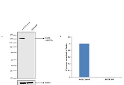
- Western blot analysis of EGFR (Fig a) was performed by loading 30 µg of A-431 Control (lane1), A-431 EGFR knockout (lane 2) whole cell extracts using Novex® NuPAGE® 10 % Bis-Tris gel, XCell SureLock™ Electrophoresis System (EI0002), Novex® Sharp Pre-Stained Protein Standard (LC5800), and iBlot® Dry Blotting System (IB21001). Proteins were transferred to a nitrocellulose membrane and blocked with 5% skim milk for 1 hour at room temperature. EGFR was detected at ~ 180 kDa using Anti-EGFR Mouse Monoclonal Antibody (Product # MA5-13269,1 µg/mL) at in 5% skim milk at 4°C overnight on a rocking platform. Goat anti-Mouse IgG (H+L) Superclonal™ Secondary Antibody HRP conjugate (Product # A28177, 1:4000 dilution) was used and chemiluminescent detection was performed using Pierce™ ECL Western Blotting Substrate (Product # 32106). Densitometric analysis of this western blot is shown in histogram (Fig b). Loss of signal in CRISPR mediated knockout (KO) confirms that antibody is specific to EGFR.

- Submitted by
- Invitrogen Antibodies (provider)
- Main image

- Experimental details
- Western blot was performed using Anti-EGFR Monoclonal Antibody (111.6) (Product # MA5-13269) and a 130 kDa band corresponding to EGFR was observed across tissues and cell lines tested. Membrane enriched extracts (30 µg lysate) of Mouse Placenta (Lane 1), Mouse Brain (Lane 2), Rat Brain (Lane 3), Mouse Liver (Lane 4), Rat Liver (Lane 5), A-431 (Lane 6) were electrophoresed using NuPAGE™ 4-12% Bis-Tris Protein Gel (Product # NP0322BOX). Resolved proteins were then transferred onto a nitrocellulose membrane (Product # IB23001) by iBlot® 2 Dry Blotting System (Product # IB21001). The blot was probed with the primary antibody (1:500 dilution) and detected by chemiluminescence with Goat anti-Mouse IgG (H+L) Superclonal™ Recombinant Secondary Antibody, HRP (Product # A28177,1:4000 dilution) using the iBright™ FL1500 Imaging System (Product # A44115). Chemiluminescent detection was performed using SuperSignal™ West Pico PLUS Chemiluminescent Substrate (Product # 34580).

Supportive validation
- Submitted by
- Invitrogen Antibodies (provider)
- Main image

- Experimental details
- Immunofluorescence analysis of EGFR was performed using 90% confluent log phase A-431 cells. The cells were fixed with 4% paraformaldehyde for 10 minutes, permeabilized with 0.1% Triton™ X-100 for 10 minutes, and blocked with 1% BSA for 1 hour at room temperature. The cells were labeled with EGFR Mouse monoclonal antibody (Product # MA5-13269) at 5 µg/mL in 0.1% BSA and incubated for 3 hours at room temperature and then labeled with Goat anti-Mouse IgG (H+L) Superclonal™ Secondary Antibody, Alexa Fluor® 488 conjugate (Product # A28175) at a dilution of 1:2000 for 45 minutes at room temperature (Panel a: green). Nuclei (Panel b: blue) were stained with SlowFade® Gold Antifade Mountant with DAPI (Product # S36938). F-actin (Panel c: red) was stained with Rhodamine Phalloidin (Product # R415, 1:300). Panel d represents the merged image showing membrane localization. Panel e shows the no primary antibody control. The images were captured at 60X magnification.

- Submitted by
- Invitrogen Antibodies (provider)
- Main image

- Experimental details
- Immunofluorescence analysis of EGFR was done on 90% confluent log phase A431 cells. The cells were fixed with 4% paraformaldehyde for 10 minutes, permeabilized with 0.1% Triton™ X-100 for 10 minutes, and blocked with 1% BSA for 1 hour at room temperature. The cells were labeled EGFR (111.6) Mouse Monoclonal Antibody (Product # MA5-13269) at 2 µg/mL in 0.1% BSA and incubated for 3 hours at room temperature and then labeled with Goat anti-Mouse IgG (H+L) Superclonal™ Secondary Antibody, Alexa Fluor® 488 conjugate (Product # A28175) at a dilution of 1:2000 for 45 minutes at room temperature (Panel a: green). Nuclei (Panel b: blue) were stained with SlowFade® Gold Antifade Mountant with DAPI (Product # S36938). F-actin (Panel c: red) was stained with Alexa Fluor® 555 Rhodamine Phalloidin (Product # R415, 1:300). Panel d is a merged image showing membranous localization. Panel e is a no primary antibody control. The images were captured at 60X magnification.

- Submitted by
- Invitrogen Antibodies (provider)
- Main image

- Experimental details
- Immunofluorescence analysis of EGFR was performed using 70% confluent log phase A-431 cells (WIld type, panels a,d), CAS9 control (panels b,e) and EGFR Knockout (panels c,f). The cells were fixed, permeabilized, and labelled with EGFR Mouse Monoclonal Antibody(Product # MA5-13269, 5 µg/mL), followed by Goat anti-Mouse IgG (H+L) Superclonal™ Secondary Antibody, Alexa Fluor® 488 conjugate (Product # A28175, 1:2000). Nuclei (blue) were stained with SlowFade® Gold Antifade Mountant with DAPI (Product # S36938) and Rhodamine Phalloidin (Product # R415, 1:300) was used for cytoskeletal F-actin (red) staining. Loss of signal was observed in EGFR Knockout cells (panel c,f) confirming specificity of the antibody to EGFR(green). The images were captured at 60X magnification.

Supportive validation
- Submitted by
- Invitrogen Antibodies (provider)
- Main image

- Experimental details
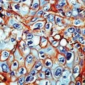
- Formalin-fixed, paraffin-embedded human lung squamous cell carcinoma stained with Epidermal Growth Factor Receptor antibody0 using peroxidase-conjugate and AEC chromogen. Note membrane staining of tumor cells.
Supportive validation
- Submitted by
- Invitrogen Antibodies (provider)
- Main image

- Experimental details
- Flow cytometry analysis of EGFR was done on A-431 cells. Cells were fixed with 70% ethanol for 10 minutes, permeabilized with 0.25% Triton™ X-100 for 20 minutes, and blocked with 5% BSA for 30 minutes at room temperature. Cells were labeled with EGFR Mouse Monoclonal Antibody (MA513269, red histogram) or with mouse isotype control (yellow histogram) at 3-5 ug/million cells in 2.5% BSA. After incubation at room temperature for 2 hours, the cells were labeled with Alexa Fluor® 488 Rabbit Anti-Mouse Secondary Antibody (A11059) at a dilution of 1:400 for 30 minutes at room temperature. The representative 10,000 cells were acquired and analyzed for each sample using an Attune® Acoustic Focusing Cytometer. The purple histogram represents unstained control cells and the green histogram represents no-primary-antibody control.

Supportive validation
- Submitted by
- Invitrogen Antibodies (provider)
- Main image
- Experimental details
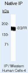
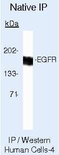
- Immunoprecipitation of Epidermal Growth Factor Receptor using Epidermal Growth Factor Receptor Monoclonal Antibody (Product # MA5-13269) on Native Human A431 Cells.
- Submitted by
- Invitrogen Antibodies (provider)
- Main image

- Experimental details
- NULL

- Submitted by
- Invitrogen Antibodies (provider)
- Main image

- Experimental details
- 8 Association between Gefitinib and OC-Exo on EGFR signaling pathway in cumulus cells. (a and b) Immunocytochemical analysis of phospho-EGFR (p-EGFR, green) and phospho-MAPK1/3 (p-MAPK1/3, green) in cumulus cells and their nuclear staining (DAPI, blue). Original magnification x100. (c) Effect of Gefitinib/OC-Exo on protein expression of p-EGFR and p-MAPK1/3 in cumulus cells. (d) Aliquots of untreated cells (control), and cells treated with 1 muM Gefitinib, with 1 muM Gefitinib and 50 ug OC-Exo, or with 50 mug OC-Exo. Cumulus cells were lysed and total cellular lysates were immunoblotted against EGFR, p-EGFR, t-MAPK1/3 (MAPK1/3), and p-MAPK1/3. Data are shown as means +- SEM. (a, b, and c) Within a column, values with different superscript letters are significantly different ( p < .05). At least three replications were performed. Control: cumulus cells cultured without Gefitinib and OC-Exo; Gefi: cumulus cells cultured with 1 muM Gefitinib; Gefi + Exo: cumulus cells cultured with 1 muM Gefitinib and 50 mug OC-Exo; Exo: cumulus cells cultured with 50 mug OC-Exo. DAPI, 4',6-diamidino-2-phenylindole; EGFR, epidermal growth factor receptor; OC-Exo, oviductal exosome; MAPK, mitogen-activated protein kinase; SEM, standard error of mean [Color figure can be viewed at wileyonlinelibrary.com]

- Submitted by
- Invitrogen Antibodies (provider)
- Main image

- Experimental details
- Fig. 2 C. pneumoniae specifically induces externalization of phosphatidylserine (PS). a Infectivity of C. pneumoniae EBs (MOI = 10) in wild-type (WT) and PS-deficient (PSA3) CHO cells ( n = 3). mean +- s.d. Infectivity in WT CHO cells was set to 100%. Mean +- s.d. b qRT PCR analysis of internalized Cpn . Triplicate samples of EBs in CHO WT and PSA3 cells (2 hpi) were pre-treated with rCPn0473 (100 µg/mL) for 1 h prior to infection (MOI = 10) ( n = 3). Mean +- s.d. c Externalization of PS by C. pneumoniae EBs early in infection. HEp-2 cells were infected with chlamydial EBs for the indicated times (MOI = 10). Externalized PS was stained with annexin-V-FLUOS prior to fixation, followed by staining with DAPI and anti-EGFR antibody. Scale bars: 2.5 um. d Externalization of PS by the indicated chlamydial species. HEp-2 cells were infected with chlamydial EBs for 1 h (MOI = 10). Externalized PS was stained as in c . Mean (triplicates) +- s.d. ( n = 3). For all panels: *** P < 0.001, ** P < 0.01 and * P < 0.05, n.s. not significant ( P > 0.05) (Student's two-sample t test). Source data are provided as a Source Data file
