 Explore
 Validate
 Learn
Learn Western blot
 ELISA
ELISAAntibody data
- Antibody Data
- Antigen structure
- References [8]
- Comments [0]
- Validations
- Western blot [1]
- Immunocytochemistry [1]
- Immunohistochemistry [1]
- Flow cytometry [1]
Submit
Validation data
Reference
Comment
Report error
- Product number
- AF231 - Provider product page

- Provider
- R&D Systems
- Product name
- Human EGFR Antibody
- Antibody type
- Polyclonal
- Description
- Antigen Affinity-purified. Detects human EGFR in ELISAs and Western blots. In sandwich ELISAs, approximately 3% cross-reactivity with recombinant mouse EGFR is observed and less than 0.1% cross-reactivity with recombinant human (rh) ErbB2 and rhErbB3 is observed.
- Reactivity
- Human
- Host
- Goat
- Conjugate
- Unconjugated
- Antigen sequence
CAA25240- Isotype
- IgG
- Vial size
- 100 ug
- Concentration
- LYOPH
- Storage
- Use a manual defrost freezer and avoid repeated freeze-thaw cycles. 12 months from date of receipt, -20 to -70 °C as supplied. 1 month, 2 to 8 °C under sterile conditions after reconstitution. 6 months, -20 to -70 °C under sterile conditions after reconstitution.
Submitted references Improved efficiency of in situ protein analysis by proximity ligation using UnFold probes.
Identification of a novel anoikis signalling pathway using the fungal virulence factor gliotoxin.
Soluble fms-like tyrosine kinase 1 promotes angiotensin II sensitivity in preeclampsia.
Ability of the Met kinase inhibitor crizotinib and new generation EGFR inhibitors to overcome resistance to EGFR inhibitors.
Human epidermal growth factor receptor (HER-1:HER-3) Fc-mediated heterodimer has broad antiproliferative activity in vitro and in human tumor xenografts.
Development and validation of sandwich ELISA microarrays with minimal assay interference.
FGFR2-amplified gastric cancer cell lines require FGFR2 and Erbb3 signaling for growth and survival.
Expression of growth factors and growth factor receptor in non-healing and healing ischaemic ulceration.
Klaesson A, Grannas K, Ebai T, Heldin J, Koos B, Leino M, Raykova D, Oelrich J, Arngården L, Söderberg O, Landegren U
Scientific reports 2018 Mar 29;8(1):5400
Scientific reports 2018 Mar 29;8(1):5400
Identification of a novel anoikis signalling pathway using the fungal virulence factor gliotoxin.
Haun F, Neumann S, Peintner L, Wieland K, Habicht J, Schwan C, Østevold K, Koczorowska MM, Biniossek M, Kist M, Busch H, Boerries M, Davis RJ, Maurer U, Schilling O, Aktories K, Borner C
Nature communications 2018 Aug 30;9(1):3524
Nature communications 2018 Aug 30;9(1):3524
Soluble fms-like tyrosine kinase 1 promotes angiotensin II sensitivity in preeclampsia.
Burke SD, Zsengellér ZK, Khankin EV, Lo AS, Rajakumar A, DuPont JJ, McCurley A, Moss ME, Zhang D, Clark CD, Wang A, Seely EW, Kang PM, Stillman IE, Jaffe IZ, Karumanchi SA
The Journal of clinical investigation 2016 Jul 1;126(7):2561-74
The Journal of clinical investigation 2016 Jul 1;126(7):2561-74
Ability of the Met kinase inhibitor crizotinib and new generation EGFR inhibitors to overcome resistance to EGFR inhibitors.
Nanjo S, Yamada T, Nishihara H, Takeuchi S, Sano T, Nakagawa T, Ishikawa D, Zhao L, Ebi H, Yasumoto K, Matsumoto K, Yano S
PloS one 2013;8(12):e84700
PloS one 2013;8(12):e84700
Human epidermal growth factor receptor (HER-1:HER-3) Fc-mediated heterodimer has broad antiproliferative activity in vitro and in human tumor xenografts.
Sarup J, Jin P, Turin L, Bai X, Beryt M, Brdlik C, Higaki JN, Jorgensen B, Lau FW, Lindley P, Liu J, Ni I, Rozzelle J, Kumari R, Watson SA, Zhang J, Shepard HM
Molecular cancer therapeutics 2008 Oct;7(10):3223-36
Molecular cancer therapeutics 2008 Oct;7(10):3223-36
Development and validation of sandwich ELISA microarrays with minimal assay interference.
Gonzalez RM, Seurynck-Servoss SL, Crowley SA, Brown M, Omenn GS, Hayes DF, Zangar RC
Journal of proteome research 2008 Jun;7(6):2406-14
Journal of proteome research 2008 Jun;7(6):2406-14
FGFR2-amplified gastric cancer cell lines require FGFR2 and Erbb3 signaling for growth and survival.
Kunii K, Davis L, Gorenstein J, Hatch H, Yashiro M, Di Bacco A, Elbi C, Lutterbach B
Cancer research 2008 Apr 1;68(7):2340-8
Cancer research 2008 Apr 1;68(7):2340-8
Expression of growth factors and growth factor receptor in non-healing and healing ischaemic ulceration.
Murphy MO, Ghosh J, Fulford P, Khwaja N, Halka AT, Carter A, Turner NJ, Walker MG
European journal of vascular and endovascular surgery : the official journal of the European Society for Vascular Surgery 2006 May;31(5):516-22
European journal of vascular and endovascular surgery : the official journal of the European Society for Vascular Surgery 2006 May;31(5):516-22
No comments: Submit comment
Supportive validation
- Submitted by
- R&D Systems (provider)
- Main image

- Experimental details
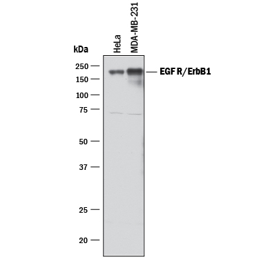
- Detection of Human EGFR by Western Blot. Western blot shows lysates of HeLa human cervical epithelial carcinoma cell line and MDA-MB-231 human breast cancer cell line. PVDF membrane was probed with 1 µg/mL of Goat Anti-Human EGFR Antigen Affinity-purified Polyclonal Antibody (Catalog # AF231) followed by HRP-conjugated Anti-Goat IgG Secondary Antibody (Catalog # HAF017). A specific band was detected for EGFR at approximately 175 kDa (as indicated). This experiment was conducted under reducing conditions and using Immunoblot Buffer Group 1.
Supportive validation
- Submitted by
- R&D Systems (provider)
- Main image

- Experimental details
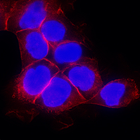
- EGFR in A431 Human Cell Line. EGFR was detected in immersion fixed A431 human epithelial carcinoma cell line using Goat Anti-Human EGFR Antigen Affinity-purified Polyclonal Antibody (Catalog # AF231) at 1 µg/mL for 3 hours at room temperature. Cells were stained using the NorthernLights™ 557-conjugated Anti-Goat IgG Secondary Antibody (red; Catalog # NL001) and counterstained with DAPI (blue). Specific staining was localized to plasma membrane. View our protocol for Fluorescent ICC Staining of Cells on Coverslips.
Supportive validation
- Submitted by
- R&D Systems (provider)
- Main image

- Experimental details
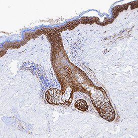
- EGFR in Human Skin. EGFR was detected in immersion fixed frozen sections of human skin using Goat Anti-Human EGFR Antigen Affinity-purified Polyclonal Antibody (Catalog # AF231) at 1 µg/mL for 1 hour at room temperature followed by incubation with the Anti-Goat IgG VisUCyte™ HRP Polymer Antibody (Catalog # VC004). Tissue was stained using DAB (brown) and counterstained with hematoxylin (blue). Specific staining was localized to plasma membrane. View our protocol for IHC Staining with VisUCyte HRP Polymer Detection Reagents.
Supportive validation
- Submitted by
- R&D Systems (provider)
- Main image
- Experimental details
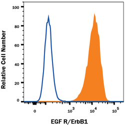
- Detection of EGFR in A431 Human Cell Line by Flow Cytometry. A431 human epithelial carcinoma cell line was stained with Goat Anti-Human EGFR Antigen Affinity-purified Polyclonal Antibody (Catalog # AF231, filled histogram) or isotype control antibody (Catalog # AB-108-C, open histogram), followed by Phycoerythrin-conjugated Anti-Goat IgG Secondary Antibody (Catalog # F0107). View our protocol for Staining Membrane-associated Proteins.
