 Explore
 Validate
 Learn
Learn Western blot
 Immunocytochemistry
 Immunohistochemistry
Antibody data
- Antibody Data
- Antigen structure
- References [2]
- Comments [0]
- Validations
- Immunocytochemistry [1]
- Immunohistochemistry [7]
Submit
Validation data
Reference
Comment
Report error
- Product number
- HPA018530 - Provider product page

- Provider
- Atlas Antibodies
- Proper citation
- Atlas Antibodies Cat#HPA018530, RRID:AB_1848044
- Product name
- Anti-EGFR
- Antibody type
- Polyclonal
- Reactivity
- Human, Mouse, Rat
- Host
- Rabbit
- Conjugate
- Unconjugated
- Antigen sequence
QQGFFSSPSTSRTPLLSSLSATSNNSTVACIDRNG
LQSCPIKEDSFLQRYSSDPTGALTEDSIDDTFLPV
PEYINQSVPKRPAGSVQNPVYHNQPLNPAPSRDPH
YQDPHSTAVGNPEYLNTVQPTCVNSTFDSPAHWAQ
KGSHQISLD- Isotype
- IgG
- Vial size
- 100 µl
- Storage
- Store at +4°C for short term storage. Long time storage is recommended at -20°C.
Submitted references Sentinel lymph node biopsy revisited: ultrasound-guided photoacoustic detection of micrometastases using molecularly targeted plasmonic nanosensors.
Proteomic screen reveals Fbw7 as a modulator of the NF-κB pathway
Luke GP, Myers JN, Emelianov SY, Sokolov KV
Cancer research 2014 Oct 1;74(19):5397-408
Cancer research 2014 Oct 1;74(19):5397-408
Proteomic screen reveals Fbw7 as a modulator of the NF-κB pathway
Arabi A, Ullah K, Branca R, Johansson J, Bandarra D, Haneklaus M, Fu J, Ariës I, Nilsson P, Den Boer M, Pokrovskaja K, Grandér D, Xiao G, Rocha S, Lehtiö J, Sangfelt O
Nature Communications 2012 July;3
Nature Communications 2012 July;3
No comments: Submit comment
Supportive validation
- Submitted by
- Atlas Antibodies (provider)
- Main image

- Experimental details
- Immunofluorescent staining of human cell line A-431 shows positivity in plasma membrane.
- Sample type
- HUMAN

Enhanced validation
Supportive validation
- Submitted by
- Atlas Antibodies (provider)
- Enhanced method
- Orthogonal validation
- Main image
- Experimental details
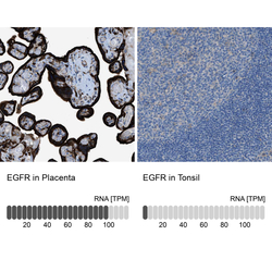
- Immunohistochemistry analysis in human placenta and tonsil tissues using HPA018530 antibody. Corresponding EGFR RNA-seq data are presented for the same tissues.
- Sample type
- HUMAN

Supportive validation
- Submitted by
- Atlas Antibodies (provider)
- Main image

- Experimental details
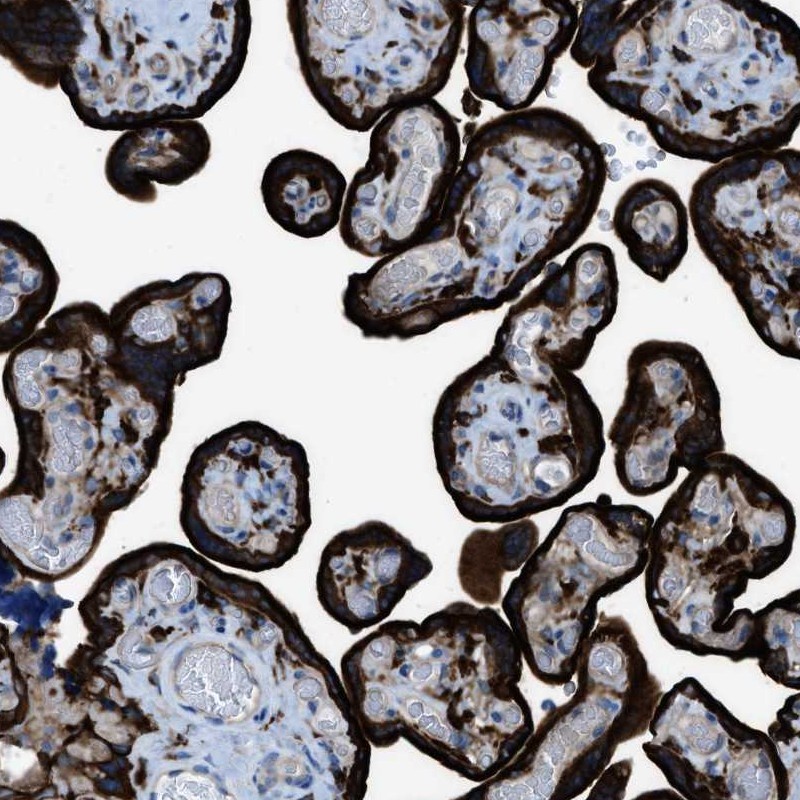
- Immunohistochemical staining of human placenta shows high expression.
- Sample type
- HUMAN
- Submitted by
- Atlas Antibodies (provider)
- Main image

- Experimental details
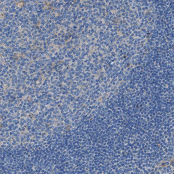
- Immunohistochemical staining of human tonsil shows low expression as expected.
- Sample type
- HUMAN

- Submitted by
- Atlas Antibodies (provider)
- Main image

- Experimental details
- Immunohistochemical staining of human cerebellum shows no positivity in Purkinje cells as expected.
- Sample type
- HUMAN

- Submitted by
- Atlas Antibodies (provider)
- Main image

- Experimental details
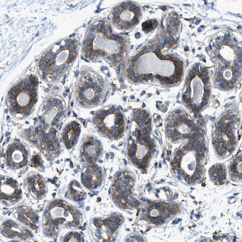
- Immunohistochemical staining of human breast shows moderate cytoplasmic positivity in glandular cells.
- Sample type
- HUMAN
- Submitted by
- Atlas Antibodies (provider)
- Main image

- Experimental details
- Immunohistochemical staining of human placenta shows strong membranous positivity in trophoblastic cells.
- Sample type
- HUMAN

- Submitted by
- Atlas Antibodies (provider)
- Main image
- Experimental details
- Immunohistochemical staining of human tonsil shows very weak positivity in germinal center cells as expected.
- Sample type
- HUMAN
