 Explore
 Validate
 Learn
Learn Western blot
 Immunocytochemistry
 Immunohistochemistry
Antibody data
- Antibody Data
- Antigen structure
- References [3]
- Comments [0]
- Validations
- Immunocytochemistry [1]
- Immunohistochemistry [7]
Submit
Validation data
Reference
Comment
Report error
- Product number
- HPA002025 - Provider product page

- Provider
- Atlas Antibodies
- Proper citation
- Atlas Antibodies Cat#HPA002025, RRID:AB_1080077
- Product name
- Anti-ERLIN2
- Antibody type
- Polyclonal
- Reactivity
- Human, Mouse, Rat
- Host
- Rabbit
- Conjugate
- Unconjugated
- Antigen sequence
KTKLLIAAQKQKVVEKEAETERKKALIEAEKVAQV
AEITYGQKVMEKETEKKISEIEDAAFLAREKAKAD
AECYTAMKIAEANKLKLTPEYLQLMKYKAIASNSK
IYFGKDIPNMFMDSAGSVSKQFEGLADKLSFGLED
EPLETATK- Isotype
- IgG
- Vial size
- 100 µl
- Storage
- Store at +4°C for short term storage. Long time storage is recommended at -20°C.
Submitted references Immunofluorescence and fluorescent-protein tagging show high correlation for protein localization in mammalian cells
is a common Luminal B breast cancer oncogene that differentially regulates luminal and basal progenitors in human mammary epithelium
Tissue profiling of the mammalian central nervous system using human antibody-based proteomics.
Stadler C, Rexhepaj E, Singan V, Murphy R, Pepperkok R, Uhlén M, Simpson J, Lundberg E
Nature Methods 2013 February;10(4):315-323
Nature Methods 2013 February;10(4):315-323
is a common Luminal B breast cancer oncogene that differentially regulates luminal and basal progenitors in human mammary epithelium
Holland D, Burleigh A, Git A, Goldgraben M, Perez-Mancera P, Chin S, Hurtado A, Bruna A, Ali H, Greenwood W, Dunning M, Samarajiwa S, Menon S, Rueda O, Lynch A, McKinney S, Ellis I, Eaves C, Carroll J, Curtis C, Aparicio S, Caldas C
EMBO Molecular Medicine 2011 March;3(3):167-180
EMBO Molecular Medicine 2011 March;3(3):167-180
Tissue profiling of the mammalian central nervous system using human antibody-based proteomics.
Mulder J, Björling E, Jonasson K, Wernérus H, Hober S, Hökfelt T, Uhlén M
Molecular & cellular proteomics : MCP 2009 Jul;8(7):1612-22
Molecular & cellular proteomics : MCP 2009 Jul;8(7):1612-22
No comments: Submit comment
Supportive validation
- Submitted by
- Atlas Antibodies (provider)
- Main image
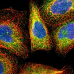
- Experimental details
- Immunofluorescent staining of human cell line A-431 shows localization to endoplasmic reticulum.
- Sample type
- HUMAN

Enhanced validation
Supportive validation
- Submitted by
- Atlas Antibodies (provider)
- Enhanced method
- Orthogonal validation
- Main image

- Experimental details
- Immunohistochemistry analysis in human kidney and skeletal muscle tissues using Anti-ERLIN2 antibody. Corresponding ERLIN2 RNA-seq data are presented for the same tissues.
- Sample type
- HUMAN

Supportive validation
- Submitted by
- Atlas Antibodies (provider)
- Main image

- Experimental details
- Immunohistochemical staining of human kidney shows high expression.
- Sample type
- HUMAN

- Submitted by
- Atlas Antibodies (provider)
- Main image

- Experimental details
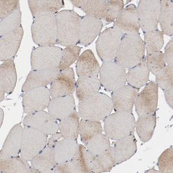
- Immunohistochemical staining of human skeletal muscle shows low expression as expected.
- Sample type
- HUMAN

- Submitted by
- Atlas Antibodies (provider)
- Main image
- Experimental details
- Immunohistochemical staining of human skeletal muscle shows very weak positivity in myocytes as expected.
- Sample type
- HUMAN

- Submitted by
- Atlas Antibodies (provider)
- Main image

- Experimental details
- Immunohistochemical staining of human kidney shows strong granular cytoplasmic positivity in cells in tubules.
- Sample type
- HUMAN

- Submitted by
- Atlas Antibodies (provider)
- Main image

- Experimental details
- Immunohistochemical staining of human Fallopian tube shows strong granular cytoplasmic positivity in glandular cells.
- Sample type
- HUMAN

- Submitted by
- Atlas Antibodies (provider)
- Main image

- Experimental details
- Immunohistochemical staining of human testis shows moderate granular cytoplasmic positivity in cells in seminiferous ducts.
- Sample type
- HUMAN
