Explore
 Validate
 Learn
Learn Immunocytochemistry
Antibody data
- Antibody Data
- Antigen structure
- References [0]
- Comments [0]
- Validations
- Immunocytochemistry [1]
- Immunohistochemistry [2]
- Other assay [2]
Submit
Validation data
Reference
Comment
Report error
- Product number
- PA5-47988 - Provider product page

- Provider
- Invitrogen Antibodies
- Product name
- FGF16 Polyclonal Antibody
- Antibody type
- Polyclonal
- Antigen
- Recombinant full-length protein
- Description
- In direct ELISAs, less than 1% cross-reactivity with recombinant human (rh) FGF acidic, rhFGF basic, rhFGF-3, -4, -5, -6, -7, -9, -10, -11, -12, -13, -17, -18, -19, -21, -23, recombinant mouse (rm) FGF-8b, and rmFGF-15 is observed.
- Concentration
- 0.2 mg/mL
No comments: Submit comment
Supportive validation
- Submitted by
- Invitrogen Antibodies (provider)
- Main image
- Experimental details
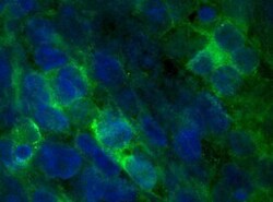
- Immunocytochemistry analysis of FGF16 in immersion fixed human induced pluripotent stem (iPS) cells differentiated into cardiomyocytes. Samples were incubated in FGF16 polyclonal antibody (Product # PA5-47988) using a dilution of 8 µg/mL for 3 hours at room temperature followed by NorthernLights™ 493-conjugated Anti-Sheep IgG Secondary Antibody (green) and counterstained with DAPI (blue). Specific staining was localized to cytoplasm.

Supportive validation
- Submitted by
- Invitrogen Antibodies (provider)
- Main image

- Experimental details
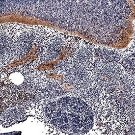
- Immunohistochemical analysis of FGF16 in perfusion fixed frozen sections of mouse embryo (13 d.p.c.). Samples were incubated in FGF16 polyclonal antibody (Product # PA5-47988) using a dilution of 15 µg/mL overnight at 4 °C. Tissue was stained using the Anti-Sheep HRP-DAB Cell & Tissue Staining Kit (brown) and counterstained with hematoxylin (blue). Specific staining was localized to roots of dorsal ganglia and spinal cord.
- Submitted by
- Invitrogen Antibodies (provider)
- Main image

- Experimental details
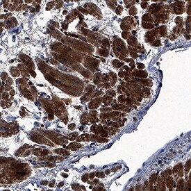
- Immunohistochemical analysis of FGF16 in immersion fixed paraffin-embedded sections of human heart. Samples were incubated in FGF16 polyclonal antibody (Product # PA5-47988) using a dilution of 3 µg/mL overnight at 4 °C. Tissue was stained using the Anti-Sheep HRP-DAB Cell & Tissue Staining Kit (brown) and counterstained with hematoxylin (blue). Specific staining was localized to cardiomyocytes.
Supportive validation
- Submitted by
- Invitrogen Antibodies (provider)
- Main image
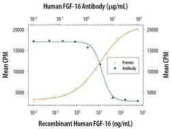
- Experimental details
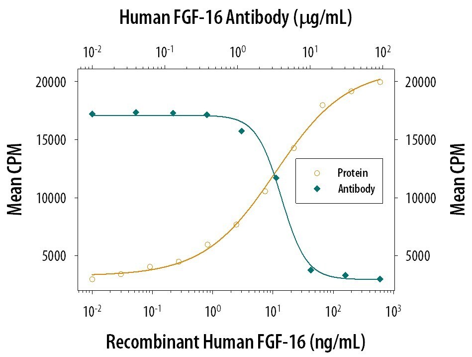
- Neutralization antibody testing demonstrates the specificity of an antibody through a correlation between antibody binding and the activity of the target. Neutralization of FGF16 is shown by the decrease in mean CPM (count per minute- counted radioactivity- measure of cell proliferation) with increasing concentrations of FGF16 Polyclonal Antibody (PA5-47988).
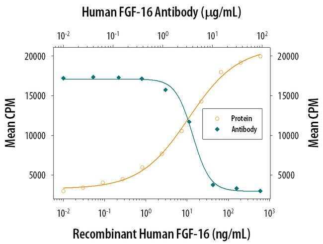
- Submitted by
- Invitrogen Antibodies (provider)
- Main image

- Experimental details
- Neutralization of FGF16 in NR6R‚3T3 mouse fibroblast cell line. Samples were incubated in FGF16 polyclonal antibody (Product # PA5-47988). Recombinant Human FGF‚16 stimulates proliferation in the NR6R‚3T3 mouse fibroblast cell line in a dose-dependent manner (orange line). Proliferation elicited by Recombinant Human FGF‚16 (100 ng/mL) is neutralized (green line) by increasing concentrations of Sheep Anti-Human/Mouse FGF‚16 Antigen Affinity-purified Polyclonal Antibody. The ND50 is typically 3-9 µg/mL.