 Explore
 Validate
 Learn
Learn Western blot
Antibody data
- Antibody Data
- Antigen structure
- References [1]
- Comments [0]
- Validations
- Western blot [2]
- Immunocytochemistry [2]
- Flow cytometry [1]
Submit
Validation data
Reference
Comment
Report error
- Product number
- 700154 - Provider product page

- Provider
- Invitrogen Antibodies
- Product name
- Phospho-FAK (Tyr861) Recombinant Rabbit Monoclonal Antibody (26H16L4)
- Antibody type
- Monoclonal
- Antigen
- Other
- Reactivity
- Human, Mouse
- Host
- Rabbit
- Isotype
- IgG
- Antibody clone number
- 26H16L4
- Vial size
- 100 µg
- Concentration
- 0.5 mg/mL
- Storage
- Store at 4°C short term. For long term storage, store at -20°C, avoiding freeze/thaw cycles.
Submitted references c-Src, Insulin-Like Growth Factor I Receptor, G-Protein-Coupled Receptor Kinases and Focal Adhesion Kinase are Enriched Into Prostate Cancer Cell Exosomes.
DeRita RM, Zerlanko B, Singh A, Lu H, Iozzo RV, Benovic JL, Languino LR
Journal of cellular biochemistry 2017 Jan;118(1):66-73
Journal of cellular biochemistry 2017 Jan;118(1):66-73
No comments: Submit comment
Supportive validation
- Submitted by
- Invitrogen Antibodies (provider)
- Main image

- Experimental details
- Western blot analysis of FAK (pY861) was performed by loading 20 µg of NIH/3T3 (lane1), NIH/3T3 treated for 10 minutes with 25 ng/mL of hPDGF (lane2) and A549 (lane3) cell lysates using Novex®NuPAGE® 4-12 % Bis-Tris gel (Product # NP0321BOX), XCell SureLock Electrophoresis System (Product # EI0002), Novex® Sharp Pre-Stained Protein Standard (Product # LC5800). Proteins were transferred to a PVDF membrane and blocked with 5 % skim milk for 1 hour at room temperature. FAK (pY861) was detected at ~125 kDa using FAK (pY861) Recombinant Rabbit Monoclonal Antibody (Product # 700154) at 1 µg-3 µg/mL in 2.5 % skim milk at 4°C overnight on a rocking platform. Goat anti-Rabbit IgG-HRP Secondary Antibody (Product # G-21234) at 1:5000 dilution was used and chemiluminescent detection was performed using Pierce™ ECL Western blotting Substrate (Product # 32106).

- Submitted by
- Invitrogen Antibodies (provider)
- Main image

- Experimental details
- Western blot analysis of Phospho-FAK/PTK2 pTyr861 in HeLa cell lysate treated with Pervanadate (30 µg/lane) using a Phospho-FAK/PTK2 pTyr861 recombinant rabbit monoclonal antibody (Product # 700154) at a dilution of 2 µg/mL. Lanes 2 and 3 show competition with the specific phosphopeptide (lane 2) or non-phosphopeptide (lane 3). NBT/BCIP was used as the substrate (Product # WB7105).

Supportive validation
- Submitted by
- Invitrogen Antibodies (provider)
- Main image

- Experimental details
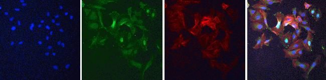
- Immunofluorescent analysis of Phospho-FAK pTyr861 in Pervanadate-treated Hela cells using a Phospho-FAK pTyr861 recombinant rabbit monoclonal antibody (Product # 700154) at a dilution of 10 µg/mL followed by detection using an Alexa Fluor 488-conjugated goat anti-rabbit secondary antibody at a dilution of 1:1000. DAPI only (blue) (top left), AF488 signal only (green) (top right), Alexa Fluor 594 Phalloidin (red) (bottom left) and the composite image (bottom right).
- Submitted by
- Invitrogen Antibodies (provider)
- Main image
- Experimental details
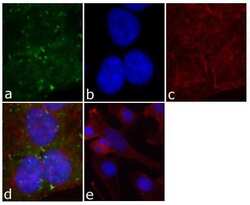
- Immunofluorescent analysis of FAK (pY861) was done on 70% confluent log phase HeLa cells. The cells were fixed with 4% paraformaldehyde for 15 minutes; permeabilized with 0.25% Triton X-100 for 10 minutes followed by blocking with 5% BSA for 1 hour at room temperature. The cells were incubated with FAK (pY861) Recombinant Rabbit Monoclonal Antibody (Product # 700154) at 2 µg-4 µg in 1% BSA and incubated for 3 hours at room temperature and then labeled with Alexa Fluor® 488 Goat anti-Rabbit IgG Secondary Antibody (Product # A-11008) at a dilution of 1:400 for 30 minutes at room temperature (Panel a: green). Nuclei (Panel b: blue) were stained with SlowFade® Gold Antifade Mountant with DAPI (Product # S36938). F-actin (Panel c: red) was stained with Alexa Fluor® 594 Phalloidin (Product # A12381). Panel d is a merged image showing cytosolic staining and punctate staining on the membrane. The images were captured at 40X magnification. Panel e shows no primary antibody control at 20x magnification.

Supportive validation
- Submitted by
- Invitrogen Antibodies (provider)
- Main image

- Experimental details
- Flow cytometry analysis of FAK [pY861] was done on A549 cells. Cells were fixed with 70% ethanol for 10 minutes, permeabilized with 0.25% Tritonª X-100 for 20 minutes, and blocked with 5% BSA for 1 hour at room temperature. Cells were labeled with ABfinityª FAK [pY861] Recombinant Rabbit Monoclonal Antibody (700154, red histogram) or with rabbit isotype control (pink histogram) at 2 µg-4 µg/million cells in 2.5% BSA. After incubation at room temperature for 2-3 hours, the cells were labeled with Alexa Fluor¨ 488 Goat Anti-Rabbit Secondary Antibody (A11008) at a dilution of 1:400 for 30 minutes at room temperature. The representative 10,000 cells were acquired and analyzed for each sample using an Attune¨ Acoustic Focusing Cytometer. The purple histogram represents unstained control cells and the green histogram represents no-primary-antibody control.
