Explore
 Validate
 Learn
Learn Western blot
 Immunoprecipitation
Antibody data
- Antibody Data
- Antigen structure
- References [1]
- Comments [0]
- Validations
- Western blot [3]
- Immunocytochemistry [2]
- Immunohistochemistry [1]
- Other assay [1]
Submit
Validation data
Reference
Comment
Report error
- Product number
- PA5-17591 - Provider product page

- Provider
- Invitrogen Antibodies
- Product name
- FAK Polyclonal Antibody
- Antibody type
- Polyclonal
- Antigen
- Synthetic peptide
- Description
- It is not recommended to aliquot this antibody. This antibody is not cross-reactive with other proteins.
- Reactivity
- Human, Mouse, Rat, Bovine, Porcine
- Host
- Rabbit
- Isotype
- IgG
- Vial size
- 100 µL
- Concentration
- 31 µg/mL
- Storage
- -20°C
Submitted references Targeting Src family kinase member Fyn by Saracatinib attenuated liver fibrosis in vitro and in vivo.
Du G, Wang J, Zhang T, Ding Q, Jia X, Zhao X, Dong J, Yang X, Lu S, Zhang C, Liu Z, Zeng Z, Safadi R, Qi R, Zhao X, Hong Z, Lu Y
Cell death & disease 2020 Feb 12;11(2):118
Cell death & disease 2020 Feb 12;11(2):118
No comments: Submit comment
Supportive validation
- Submitted by
- Invitrogen Antibodies (provider)
- Main image

- Experimental details
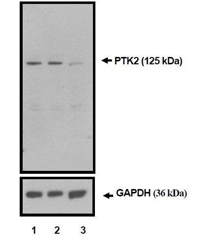
- Western blot analysis of PTK2 was performed with 10 µg of A549 cells transfected with Transfection Reagent alone (Lane 1), 100nM Non-Targeting control siRNA (Lane 2), or 100nM siRNA against PTK2 (Lane 3). Proteins were resolved using a NuPAGE® Novex 4-12% Bis-Tris Gel (Product # NP0322BOX), XCell SureLock™ Electrophoresis System (Product # EI0002), and a protein size ladder. Proteins were wet transferred to a Pierce Nitrocellulose Membrane (Product # 88025) OR Pierce PVDF Membrane (Product # 88518) and blocked with Pierce Starting Block T20 (PBS) Blocking Buffer (Product # 37539) for 1 hour at room temperature. PTK2 was detected at ~ 125 kDa using PTK2 Rabbit polyclonal antibody (Product # PA5-17591) diluted in Pierce Starting Block T20 (PBS) Blocking Buffer 4°C overnight on a rocking platform. Pierce Goat Anti-Rabbit (Product # 31461) HRP-Conjugated Antibodies at a 1:2500 dilution were used and chemiluminescent detection was performed using Pierce Supersignal West Dura Maximum Sensitivity Substrate (Product # 37071). Relative density of the bands normalized to GAPDH (36 kDa). PTK2 Antibody (Product # PA5-17591) confirms silencing of PTK2 expression.
- Submitted by
- Invitrogen Antibodies (provider)
- Main image
- Experimental details
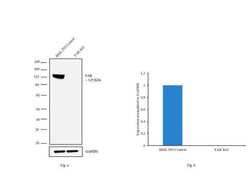
- Western blot analysis of FAK (Fig. a) was performed by loading 20 µg of HEK-293 Control (lane 1), HEK-293 FAK knockout (lane 2) whole cell extracts. FAK was detected at 125 kDa using FAK rabbit polyclonal Antibody (Product # PA5-17591, 1:1000 dilution) and Goat anti-Rabbit IgG (H+L) Superclonal™ Secondary Antibody HRP conjugate (Product # A27036, 1:4000 dilution). Densitometric analysis of this western blot is shown in histogram (Fig. b). Loss of signal in CRISPR mediated knockout (KO) confirms that antibody is specific to FAK.

- Submitted by
- Invitrogen Antibodies (provider)
- Main image

- Experimental details
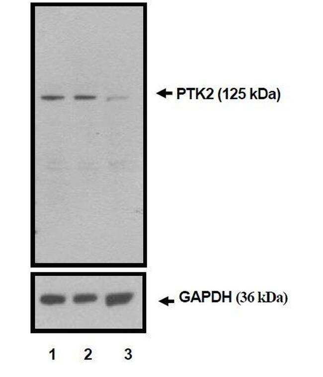
- Western blot analysis of PTK2 was performed with 10 µg of A549 cells transfected with Transfection Reagent alone (Lane 1), 100nM Non-Targeting control siRNA (Lane 2), or 100nM siRNA against PTK2 (Lane 3). Proteins were resolved using a NuPAGE® Novex 4-12% Bis-Tris Gel (Product # NP0322BOX), XCell SureLock™ Electrophoresis System (Product # EI0002), and a protein size ladder. Proteins were wet transferred to a Pierce Nitrocellulose Membrane (Product # 88025) OR Pierce PVDF Membrane (Product # 88518) and blocked with Pierce Starting Block T20 (PBS) Blocking Buffer (Product # 37539) for 1 hour at room temperature. PTK2 was detected at ~ 125 kDa using PTK2 Rabbit polyclonal antibody (Product # PA5-17591) diluted in Pierce Starting Block T20 (PBS) Blocking Buffer 4°C overnight on a rocking platform. Pierce Goat Anti-Rabbit (Product # 31461) HRP-Conjugated Antibodies at a 1:2500 dilution were used and chemiluminescent detection was performed using Pierce Supersignal West Dura Maximum Sensitivity Substrate (Product # 37071). Relative density of the bands normalized to GAPDH (36 kDa). PTK2 Antibody (Product # PA5-17591) confirms silencing of PTK2 expression.
Supportive validation
- Submitted by
- Invitrogen Antibodies (provider)
- Main image
- Experimental details
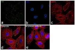
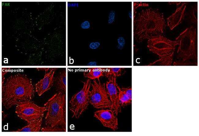
- Immunofluorescence analysis of FAK was performed using 70% confluent log phase A549 cells. The cells were fixed with 4% paraformaldehyde for 10 minutes, permeabilized with 0.1% Triton™ X-100 for 10 minutes, and blocked with 2% BSA for 1 hour at room temperature. The cells were labeled with FAK Polyclonal Antibody (Product # PA5-17591) at 1:100 dilution in 0.1% BSA and incubated overnight at 4 degree and then labeled with Goat anti-Rabbit IgG (H+L) Superclonal™ Secondary Antibody, Alexa Fluor® 488 conjugate (Product # A27034) at a dilution of 1:2000 for 45 minutes at room temperature (Panel a: green). Nuclei (Panel b: blue) were stained with SlowFade® Gold Antifade Mountant with DAPI (Product # S36938). F-actin (Panel c: red) was stained with Rhodamine Phalloidin (Product # R415, 1:300). Panel d represents the merged image showing localization at focal adhesions. Panel e represents control cells with no primary antibody to assess background. The images were captured at 60X magnification.
- Submitted by
- Invitrogen Antibodies (provider)
- Main image

- Experimental details
- Knockdown of FAK was achieved by transfecting HeLa cells with FAK specific siRNA (Silencer® select Cat # s5007*). Immunofluorescence analysis was performed on HeLa cells (untransfected, panel a,d), transfected with non-specific scrambled siRNA (panels b,e) and transfected with FAK specific siRNA (panel c,f). Cells were fixed, permeabilized, and labelled with FAK polyclonal Antibody (Product # PA5-17591, 5 µg/mL), followed by Goat anti-Rabbit IgG (H+L) Superclonal™ Secondary Antibody, Alexa Fluor® 488 conjugate (Product # A27034, 1:2000). Nuclei (blue) were stained using SlowFade® Gold Antifade Mountant with DAPI (Product # S36938), and Rhodamine Phalloidin (Product # R415, 1:300) was used for cytoskeletal F-actin (red) staining. Loss of signal was observed upon siRNA mediated knockdown (panel c,f) confirming specificity of the antibody to FAK (green). The images were captured at 60X magnification.

Supportive validation
- Submitted by
- Invitrogen Antibodies (provider)
- Main image
- Experimental details
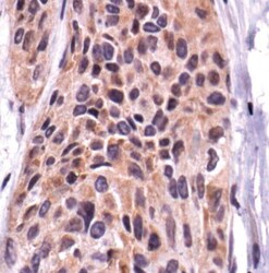
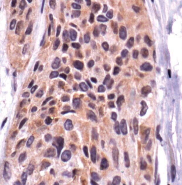
- Immunohistochemical analysis of FAK using a polyclonal antibody (Product # PA5-17591).
Supportive validation
- Submitted by
- Invitrogen Antibodies (provider)
- Main image
- Experimental details
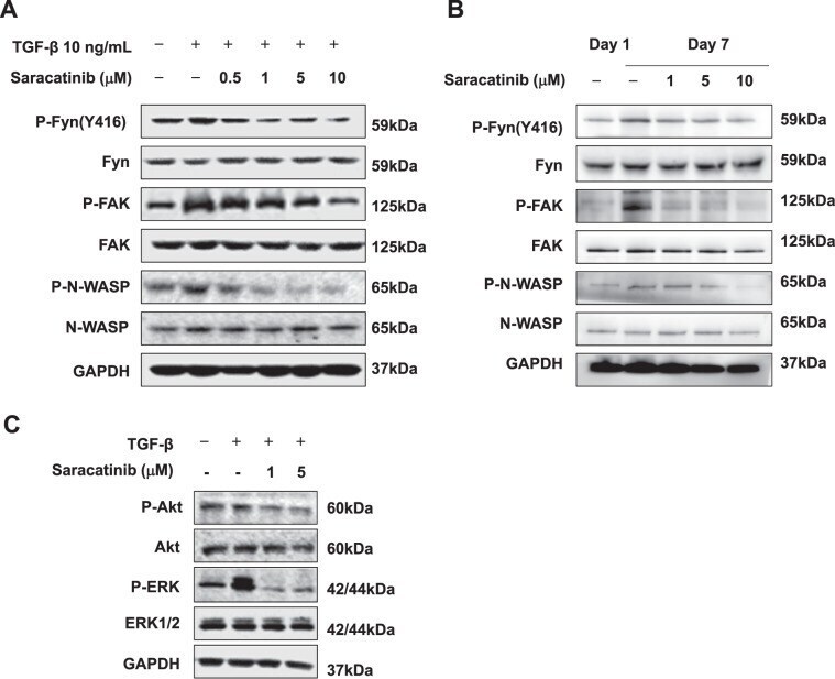
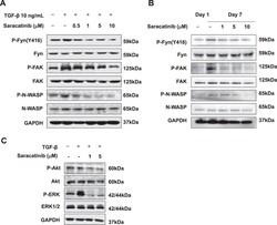
- Fig. 6 Saracatinib suppressed Fyn/FAK/N-WASP pathway. Western blot analysis on the expression of phosphorylated Fyn, FAK, and N-WASP in LX-2 cells ( a ) and mouse primary HSCs ( b ) with Saracatinib, following treatment with 10 ng/mL TGF-beta. c Western blot analysis showed that Saracatinib suppressed the expression of phosphorylated Akt and ERK in LX-2 cells treated with 10 ng/mL TGF-beta. Data are from a representative experiment that was repeated three times.