 Explore
 Validate
 Learn
Learn Western blot
Antibody data
- Antibody Data
- Antigen structure
- References [7]
- Comments [0]
- Validations
- Western blot [1]
- Immunocytochemistry [3]
- Immunohistochemistry [2]
- Flow cytometry [2]
- Other assay [1]
Submit
Validation data
Reference
Comment
Report error
- Product number
- 700047 - Provider product page

- Provider
- Invitrogen Antibodies
- Product name
- Phospho-SMAD1/SMAD5 (Ser463, Ser465) Recombinant Rabbit Monoclonal Antibody (31H14L11)
- Antibody type
- Monoclonal
- Antigen
- Synthetic peptide
- Description
- This antibody is predicted to react with mouse, primate, rat, Rhesus monkey , sea urchin, ovine, porcine, Xenopus and zebrafish based on sequence homology.
- Antibody clone number
- 31H14L11
- Concentration
- 0.5 mg/mL
Submitted references Targeting local lymphatics to ameliorate heterotopic ossification via FGFR3-BMPR1a pathway.
Fibroblast-derived Gremlin1 localises to epithelial cells at the base of the intestinal crypt.
CRIM1 is necessary for coronary vascular endothelial cell development and homeostasis.
Enhancer of Zeste Homolog 2 Inhibition Stimulates Bone Formation and Mitigates Bone Loss Caused by Ovariectomy in Skeletally Mature Mice.
Crim1 has cell-autonomous and paracrine roles during embryonic heart development.
ROBO2 restricts the nephrogenic field and regulates Wolffian duct-nephrogenic cord separation.
Essential role of Bmp signaling and its positive feedback loop in the early cell fate evolution of chordates.
Zhang D, Huang J, Sun X, Chen H, Huang S, Yang J, Du X, Tan Q, Luo F, Zhang R, Zhou S, Jiang W, Ni Z, Wang Z, Jin M, Xu M, Li F, Chen L, Liu M, Su N, Luo X, Yin L, Zhu Y, Feng JQ, Chen D, Qi H, Chen L, Xie Y
Nature communications 2021 Jul 19;12(1):4391
Nature communications 2021 Jul 19;12(1):4391
Fibroblast-derived Gremlin1 localises to epithelial cells at the base of the intestinal crypt.
Dutton LR, Hoare OP, McCorry AMB, Redmond KL, Adam NE, Canamara S, Bingham V, Mullan PB, Lawler M, Dunne PD, Brazil DP
Oncotarget 2019 Jul 23;10(45):4630-4639
Oncotarget 2019 Jul 23;10(45):4630-4639
CRIM1 is necessary for coronary vascular endothelial cell development and homeostasis.
Iyer S, Chhabra Y, Harvey TJ, Wang R, Chiu HS, Smith AG, Thomas WG, Pennisi DJ, Piper M
Journal of molecular histology 2017 Feb;48(1):53-61
Journal of molecular histology 2017 Feb;48(1):53-61
Enhancer of Zeste Homolog 2 Inhibition Stimulates Bone Formation and Mitigates Bone Loss Caused by Ovariectomy in Skeletally Mature Mice.
Dudakovic A, Camilleri ET, Riester SM, Paradise CR, Gluscevic M, O'Toole TM, Thaler R, Evans JM, Yan H, Subramaniam M, Hawse JR, Stein GS, Montecino MA, McGee-Lawrence ME, Westendorf JJ, van Wijnen AJ
The Journal of biological chemistry 2016 Nov 18;291(47):24594-24606
The Journal of biological chemistry 2016 Nov 18;291(47):24594-24606
Crim1 has cell-autonomous and paracrine roles during embryonic heart development.
Iyer S, Chou FY, Wang R, Chiu HS, Raju VK, Little MH, Thomas WG, Piper M, Pennisi DJ
Scientific reports 2016 Jan 29;6:19832
Scientific reports 2016 Jan 29;6:19832
ROBO2 restricts the nephrogenic field and regulates Wolffian duct-nephrogenic cord separation.
Wainwright EN, Wilhelm D, Combes AN, Little MH, Koopman P
Developmental biology 2015 Aug 15;404(2):88-102
Developmental biology 2015 Aug 15;404(2):88-102
Essential role of Bmp signaling and its positive feedback loop in the early cell fate evolution of chordates.
Kozmikova I, Candiani S, Fabian P, Gurska D, Kozmik Z
Developmental biology 2013 Oct 15;382(2):538-54
Developmental biology 2013 Oct 15;382(2):538-54
No comments: Submit comment
Supportive validation
- Submitted by
- Invitrogen Antibodies (provider)
- Main image
- Experimental details
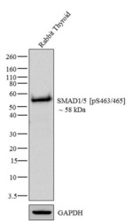
- Western blot analysis of SMAD1/5 (pS463/465) was performed by loading 20 µg of Rabbit Thyroid tissue lysate using Novex®NuPAGE®4-12% Bis-Tris gel (Product # NP0321BOX), XCell SureLock Electrophoresis System (Product # EI0002), Novex® Sharp Pre-Stained Protein Standard (Product # LC5800), and iBlot® Dry Blotting System (Product # IB21001). Proteins were transferred to a nitrocellulose membrane and blocked with 5% skim milk for 1 hour at room temperature. SMAD1/5 (pS463/465) was detected at ~58 kDa using SMAD1/5 (pS463/465) Recombinant Rabbit Monoclonal Antibody (Product # 700047) at 1-2 µg/mL in 2.5% skim milk at 4°C overnight on a rocking platform. Goat anti-Rabbit IgG - HRP Secondary Antibody (Product # G-21234) at 1:5000 dilution was used and chemiluminescent detection was performed using Pierce™ ECL Western blotting Substrate (Product # 32106).

Supportive validation
- Submitted by
- Invitrogen Antibodies (provider)
- Main image

- Experimental details
- Immunofluorescent analysis of Phospho-SMAD1/5 pSer463/465 in HeLa cells using a Phospho-SMAD1/5 pSer463/465 recombinant rabbit monoclonal antibody (Product # 700047) at a dilution of 2.5 µg/mL in the absence of peptide (top left) and presence of phosphopeptide used as immunogen (top right) or non-phosphopeptide (bottom left), followed by detection using an Alexa Fluor 488-conjugated goat anti-rabbit secondary antibody at a dilution of 1:1000. Actin was stained with Alexa Fluor 568 phalloidin (Product # A12380).

- Submitted by
- Invitrogen Antibodies (provider)
- Main image

- Experimental details
- Immunofluorescence analysis of SMAD1/5 (pS463/465) was done on 70% confluent log phase HeLa cells. The cells were fixed with 4% paraformaldehyde for 15 minutes, permeabilized with 0.25% Triton X-100 for 10 minutes, and blocked with 5% BSA for 1 hour at room temperature. The cells were labeled with SMAD1/5 (pS463/465) Recombinant Rabbit Monoclonal Antibody (Product # 700047) at 2 µg/mL in 1% BSA and incubated for 3 hours at room temperature and then labeled with Alexa Fluor 488 Goat anti-Rabbit IgG Secondary Antibody (Product # A-11008) at a dilution of 1:400 for 30 minutes at room temperature (Panel a: green). Nuclei (Panel b: blue) were stained with SlowFade® Gold Antifade Mountant DAPI (Product # S36938). F-actin (Panel c: red) was stained with Alexa Fluor 594 Phalloidin (Product # A12381). Panel d is a merged image showing nuclear localization. Panel e shows no primary antibody control. The images were captured at 20X magnification.

Supportive validation
- Submitted by
- Invitrogen Antibodies (provider)
- Main image
- Experimental details
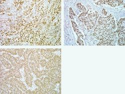
- Immunohistochemistry analysis of Phospho-SMAD1/5 pSer463/465 in formalin-fixed, paraffin-embedded human lung (top left), breast (top right) and thyroid carcimona (bottom) using a Phospho-SMAD1/5 pSer463/465 monoclonal antibody (Product # 700047) at a dilution of 5 µg/mL. Tissues were pretreated with EDTA and staining was visualized using DAB. Images were taken at a magnification of 20x. Results show nuclear staining in tumor cells.

- Submitted by
- Invitrogen Antibodies (provider)
- Main image
- Experimental details
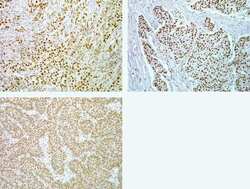
- Immunohistochemistry analysis of Phospho-SMAD1/5 pSer463/465 in formalin-fixed, paraffin-embedded human lung (top left), breast (top right) and thyroid carcimona (bottom) using a Phospho-SMAD1/5 pSer463/465 monoclonal antibody (Product # 700047) at a dilution of 5 µg/mL. Tissues were pretreated with EDTA and staining was visualized using DAB. Images were taken at a magnification of 20x. Results show nuclear staining in tumor cells.

Supportive validation
- Submitted by
- Invitrogen Antibodies (provider)
- Main image

- Experimental details
- Flow cytometry analysis of Smad1/5 [pS463/465] was done on serum starved HeLa cells. Cells were fixed with 70% ethanol for 10 minutes, permeabilized with 0.25% Triton™ X-100 for 20 minutes, and blocked with 5% BSA for 30 minutes at room temperature. Cells were labeled with ABfinity™ Smad1/5 [pS463/465] Recombinant Rabbit Monoclonal Antibody (700047, red histogram) or with rabbit isotype control (pink histogram) at 3-5 µg/million cells in 2.5% BSA. After incubation at room temperature for 2 hours, the cells were labeled with Alexa Fluor® 488 Goat Anti-Rabbit Secondary Antibody (A11008) at a dilution of 1:400 for 30 minutes at room temperature. The representative 10,000 cells were acquired and analyzed for each sample using an Attune® Acoustic Focusing Cytometer. The purple histogram represents unstained control cells and the green histogram represents no-primary-antibody control.

- Submitted by
- Invitrogen Antibodies (provider)
- Main image

- Experimental details
- Flow cytometry analysis of Phospho-SMAD1/5 pSer463/465 in Jurkat cells stimulated with BMP-4 (black) or unstimulated (gray) using a Phospho-SMAD1/5 pSer463/465 recombinant rabbit monoclonal antibody (Product # 700047) at a dilution of 0.5 µg. Cells were fixed and permeabilized using FIX & PERM (Product # GAS-004) reagent, and detection was performed using an Alexa Fluor 488 goat anti-rabbit IgG. Pre-incubation with the immunogenic peptide decreased the signal (red).

Supportive validation
- Submitted by
- Invitrogen Antibodies (provider)
- Main image

- Experimental details
- NULL
