 Explore
 Validate
 Learn
Learn Western blot
Antibody data
- Antibody Data
- Antigen structure
- References [0]
- Comments [0]
- Validations
- Western blot [1]
- Immunocytochemistry [1]
- Chromatin Immunoprecipitation [1]
Submit
Validation data
Reference
Comment
Report error
- Product number
- 703052 - Provider product page

- Provider
- Invitrogen Antibodies
- Product name
- C17orf96 Recombinant Rabbit Monoclonal Antibody (5H7L8)
- Antibody type
- Monoclonal
- Antigen
- Synthetic peptide
- Description
- This antibody is predicted to react with Monkey, Pig, Cat, Dog.
- Antibody clone number
- 5H7L8
- Concentration
- 0.5 mg/mL
No comments: Submit comment
Supportive validation
- Submitted by
- Invitrogen Antibodies (provider)
- Main image

- Experimental details
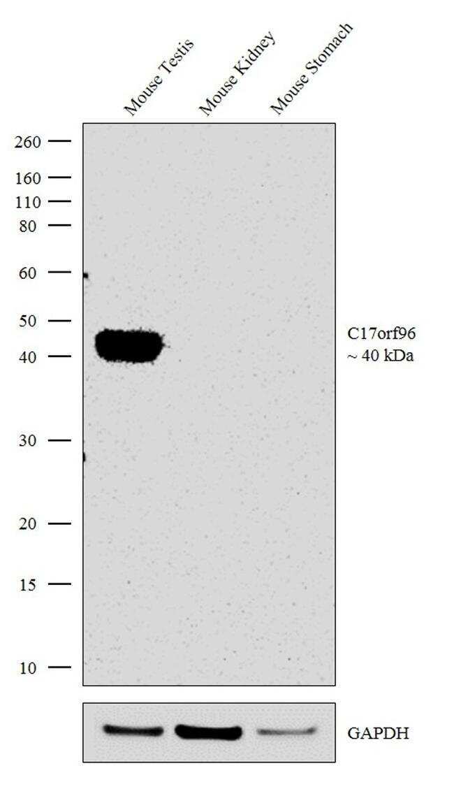
- Western blot analysis was performed on tissue extracts (30 µg lysate) of Mouse Testis (Lane 1), Mouse Kidney (Lane 2) and Mouse Stomach (Lane 3). The blot was probed with Anti-C17orf96 Recombinant Rabbit Monoclonal Antibody (Product # 703052, 1:500 dilution) and detected by chemiluminescence using Goat anti-Rabbit IgG (H+L) Superclonal™ Secondary Antibody, HRP conjugate (Product # A27036, 1:4000 dilution). A ~40 kDa band corresponding to C17orf96 was observed in the relevant tissues tested.
Supportive validation
- Submitted by
- Invitrogen Antibodies (provider)
- Main image

- Experimental details
- For immunofluorescence analysis, HEK-293 cells were fixed and permeabilized for detection of endogenous C17orf96 using Anti-C17orf96 Recombinant Rabbit Monoclonal Antibody (Product # 703052, 1:100 dilution) and labeled with Goat anti-Rabbit IgG (H+L) Superclonal™ Secondary Antibody, Alexa Fluor® 488 conjugate (Product # A27034, 1:2000). Panel a) shows representative cells that were stained for detection and localization of C17orf96 protein (green), Panel b) is stained for nuclei (blue) using ProLong™ Diamond Antifade Mountant with DAPI (Product # P36962). Panel c) represents cytoskeletal F-actin staining using Rhodamine Phalloidin (Product # R415, 1:300). Panel d) is a composite image of Panels a, b and c clearly demonstrating nuclear localization of C17orf96. Panel e) represents control cells with no primary antibody to assess background. The images were captured at 60X magnification.

Supportive validation
- Submitted by
- Invitrogen Antibodies (provider)
- Main image

- Experimental details
- Enrichment of endogenous C17ORF96 protein at specific gene loci using Anti-C17ORF96 Antibody: Chromatin Immunoprecipitation (ChIP) was performed using Anti-C17ORF96 Recombinant Rabbit Monoclonal Antibody (Product # 703052, 5 µg) on sheared chromatin from 2 million HEK-293 cells using the MAGnify ChIP System kit (Product # 492024). Normal Rabbit IgG was used as a negative IP control. The purified DNA was analyzed by qPCR with PCR primer pairs over Promoters of HOXA9, MYT1, SOX2 and SAT2 satellite repeats . Data is presented as fold enrichment of the antibody signal versus the negative control IgG using the comparative CT method.
