Explore
 Validate
 Learn
Learn Western blot
Antibody data
- Antibody Data
- Antigen structure
- References [2]
- Comments [0]
- Validations
- Western blot [6]
- Immunocytochemistry [2]
- Immunohistochemistry [1]
- Other assay [2]
Submit
Validation data
Reference
Comment
Report error
- Product number
- PA5-40127 - Provider product page

- Provider
- Invitrogen Antibodies
- Product name
- STX17 Polyclonal Antibody
- Antibody type
- Polyclonal
- Antigen
- Recombinant full-length protein
- Description
- Recommended positive controls: 293T, A431, HeLa, and HepG2 cells.
- Concentration
- 1.33 mg/mL
Submitted references The CD36 Ligand-Promoted Autophagy Protects Retinal Pigment Epithelial Cells from Oxidative Stress.
STX17 dynamically regulated by Fis1 induces mitophagy via hierarchical macroautophagic mechanism.
Dorion MF, Mulumba M, Kasai S, Itoh K, Lubell WD, Ong H
Oxidative medicine and cellular longevity 2021;2021:6691402
Oxidative medicine and cellular longevity 2021;2021:6691402
STX17 dynamically regulated by Fis1 induces mitophagy via hierarchical macroautophagic mechanism.
Xian H, Yang Q, Xiao L, Shen HM, Liou YC
Nature communications 2019 May 3;10(1):2059
Nature communications 2019 May 3;10(1):2059
No comments: Submit comment
Supportive validation
- Submitted by
- Invitrogen Antibodies (provider)
- Main image

- Experimental details
- Western blot analysis of Syntaxin-17 in various whole cell extracts (30 µg). Samples were separated by 12% SDS-PAGE and the membrane was probed with a Syntaxin-17 polyclonal antibody (Product # PA5-40127) at a dilution of 1:1000.

- Submitted by
- Invitrogen Antibodies (provider)
- Main image

- Experimental details
- Western Blot analysis of STX17 was performed by separating 30 µg of various whole cell extracts by 12% SDS-PAGE. Proteins were transferred to a membrane and probed with a STX17 Polyclonal Antibody (Product # PA5-40127) at a dilution of 1:1000 and a HRP-conjugated anti-rabbit IgG secondary antibody.

- Submitted by
- Invitrogen Antibodies (provider)
- Main image

- Experimental details
- Western Blot using STX17 Polyclonal Antibody (Product # PA5-40127). Various whole cell extracts (30 µg) were separated by 12% SDS-PAGE, and the membrane was blotted with STX17 Polyclonal Antibody (Product # PA5-40127) diluted at 1:1,000. The HRP-conjugated anti-rabbit IgG antibody was used to detect the primary antibody.

- Submitted by
- Invitrogen Antibodies (provider)
- Main image

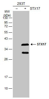
- Experimental details
- Western Blot analysis of STX17 was performed by separating 30 µg of non-transfected (–) and transfected (+) 293T whole cell extracts by 12% SDS-PAGE. Proteins were transferred to a membrane and probed with a STX17 Polyclonal Antibody (Product # PA5-40127) at a dilution of 1:5000. The HRP-conjugated anti-rabbit IgG antibody was used to detect the primary antibody.
- Submitted by
- Invitrogen Antibodies (provider)
- Main image

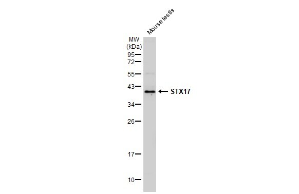
- Experimental details
- Western Blot using STX17 Polyclonal Antibody (Product # PA5-40127). Mouse tissue extract (50 µg) was separated by 12% SDS-PAGE, and the membrane was blotted with STX17 Polyclonal Antibody (Product # PA5-40127) diluted at 1:1,000. The HRP-conjugated anti-rabbit IgG antibody was used to detect the primary antibody, and the signal was developed with Trident ECL plus-Enhanced.
- Submitted by
- Invitrogen Antibodies (provider)
- Main image

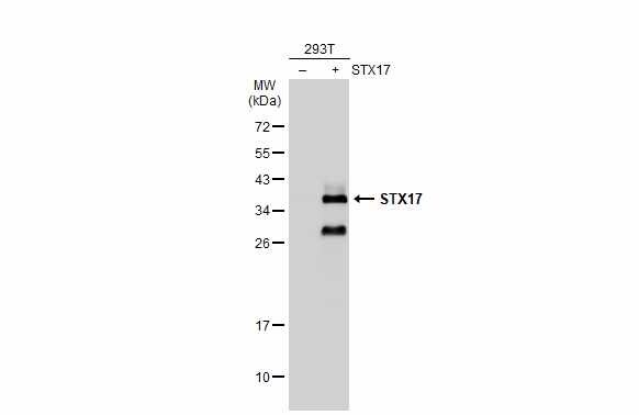
- Experimental details
- Western Blot using STX17 Polyclonal Antibody (Product # PA5-40127). Non-transfected (–) and transfected (+) 293T whole cell extracts (30 µg) were separated by 12% SDS-PAGE, and the membrane was blotted with STX17 Polyclonal Antibody (Product # PA5-40127) diluted at 1:5,000. The HRP-conjugated anti-rabbit IgG antibody was used to detect the primary antibody.
Supportive validation
- Submitted by
- Invitrogen Antibodies (provider)
- Main image

- Experimental details
- Immunocytochemistry-Immunofluorescence analysis of STX17 was performed in DIV9 rat E18 primary cortical neurons fixed in 4% paraformaldehyde at RT for 15 min. Green: STX17 Polyclonal Antibody (Product # PA5-40127) diluted at 1:500. Red: beta Tubulin 3/ Tuj1, stained by beta Tubulin 3/ Tuj1 antibody. Blue: Fluoroshield with DAPI.

- Submitted by
- Invitrogen Antibodies (provider)
- Main image
- Experimental details
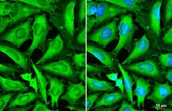
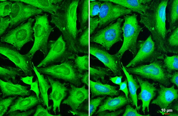
- STX17 Polyclonal Antibody detects STX17 protein by immunofluorescent analysis. Sample: HeLa cells were fixed in 4% paraformaldehyde at RT for 15 min. Green: STX17 stained by STX17 Polyclonal Antibody (Product # PA5-40127) diluted at 1:500. Blue: Fluoroshield with DAPI . Scale bar= 10 µm.
Supportive validation
- Submitted by
- Invitrogen Antibodies (provider)
- Main image

- Experimental details
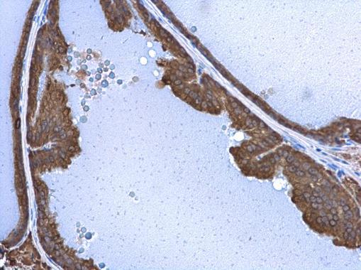
- STX17 Polyclonal Antibody detects STX17 protein at cytoplasm in mouse prostate by immunohistochemical analysis. Sample: Paraffin-embedded mouse prostate. STX17 Polyclonal Antibody (Product # PA5-40127) diluted at 1:500. Antigen Retrieval: Citrate buffer, pH 6.0, 15 min.
Supportive validation
- Submitted by
- Invitrogen Antibodies (provider)
- Main image
- Experimental details
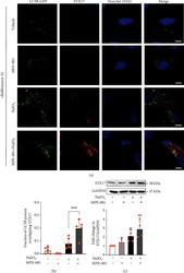
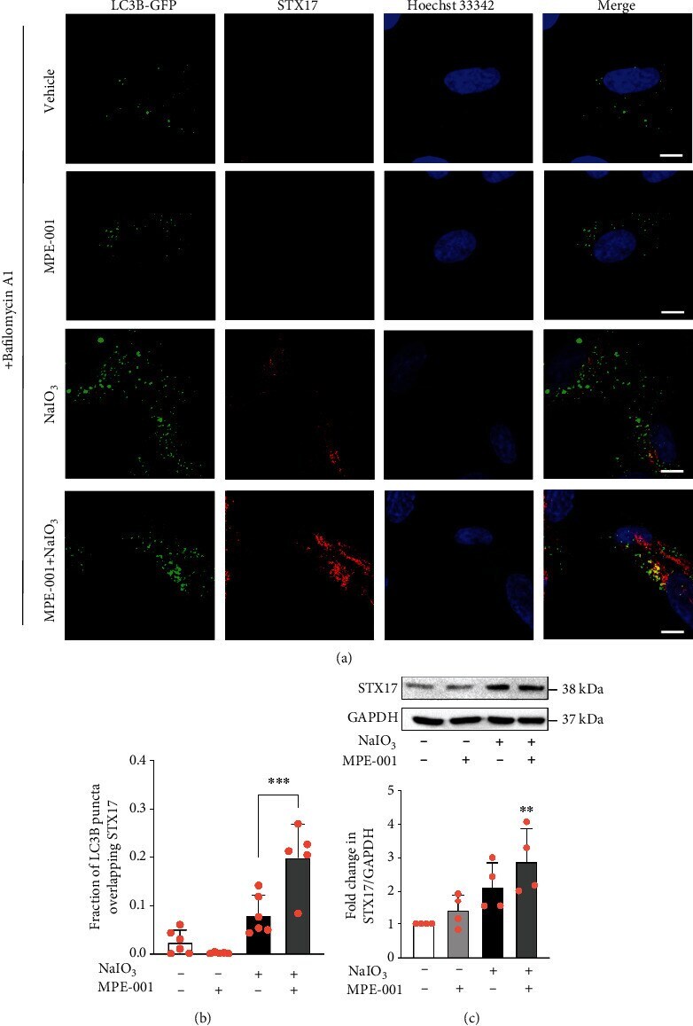
- Figure 5 MPE-001 increased the recruitment of STX17 to autophagosomes in NaIO 3 -treated cells. hTERT RPE-1 cells were pretreated with 1 mu M MPE-001 for 2 h and then exposed to 12.5 mM NaIO 3 for 4 h. (a) Representative images of LC3B-GFP-transfected cells immunostained for STX17 following treatments in the presence of 10 nM bafilomycin A1 (scale bar = 10 mu m). The same gamma correction was applied to all images. (b) Fraction of LC3B puncta that overlap STX17 staining. Manders' colocalization coefficient analysis was carried out across a series of 5 to 6 images of 6 to 18 cells each. Mean +- SD, *** p < 0.001. (c) Immunoblot of STX17 and GAPDH (upper) and relative quantification of STX17/GAPDH (lower). n = 4, mean +- SD, ns: nonsignificant, and ** p < 0.01 vs vehicle.
- Submitted by
- Invitrogen Antibodies (provider)
- Main image

- Experimental details
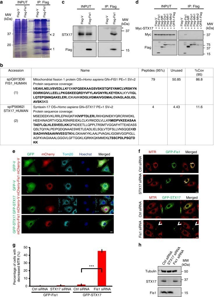
- Fig. 1 Mitochondrial fission 1 protein (Fis1) and syntaxin 17 (STX17) interact and partially colocalize. a , b HeLa cells were transfected with Flag-tagged vector or Fis1. After 24 h, cells were collected for immunoprecipitation (IP) with anti-Flag beads. Coomassie blue staining was used to visualize bands 1 and 2 ( a ). Results for mass spectrometry analysis of band 1 and 2 are summarized ( b ). c Cells treated as in a were extracted. Anti-Flag immunoprecipitates were separated by sodium dodecyl sulfate-polyacrylamide gel electrophoresis (SDS-PAGE) and immunoblotted for STX17 and Flag. Asterisk indicates a non-specific band. d HEK293T cells were co-transfected with Myc-tagged STX17 and Flag-tagged plasmids as indicated. Cells were solubilized for IP with anti-Flag and analyzed with Myc and Flag antibodies respectively. e HeLa cells were transfected with green fluorescent protein (GFP)-tagged vector or STX17 (green) and mCherry-tagged vector or plasmid encoding Fis1 (red) for 24 h. Cells were fixed and stained with anti-Tom20 (cyan). Hoechst, blue. Scale bar, 10 um. f HeLa cells were treated with the indicated small interfering RNA (siRNA) for 24 h before transfecting with GFP-tagged Fis1 (green) or GFP-STX17 (green) for further 24 h. Representative confocal images of live cells are shown. Mitochondrial morphology was visualized using MitoTracker Red (MTR, red). Scale bar, 10 um. White arrowhead indicates cells with decreased MTR. g Quantification of cells with