Explore
 Validate
 Learn
Learn Western blot
Antibody data
- Antibody Data
- Antigen structure
- References [2]
- Comments [0]
- Validations
- Western blot [1]
- Immunocytochemistry [1]
- Other assay [1]
Submit
Validation data
Reference
Comment
Report error
- Product number
- MA5-14220 - Provider product page

- Provider
- Invitrogen Antibodies
- Product name
- MMP9 Monoclonal Antibody (IA5)
- Antibody type
- Monoclonal
- Antigen
- Other
- Description
- MA5-14220 targets MMP-9 (92kDa Collagenase IV) in ICC/IF, IP, and WB applications and shows reactivity with Human samples. The MA5-14220 immunogen is human native 92kDa MMP-9.
- Reactivity
- Human
- Host
- Mouse
- Isotype
- IgG
- Antibody clone number
- IA5
- Vial size
- 500 µL
- Concentration
- 0.2 mg/mL
- Storage
- 4° C
Submitted references MicroRNA-27a Inhibits Cell Migration and Invasion of Fibroblast-Like Synoviocytes by Targeting Follistatin-Like Protein 1 in Rheumatoid Arthritis.
Inflammatory infiltrates and neovessels are relevant sources of MMPs in abdominal aortic aneurysm wall.
Shi DL, Shi GR, Xie J, Du XZ, Yang H
Molecules and cells 2016 Aug 31;39(8):611-8
Molecules and cells 2016 Aug 31;39(8):611-8
Inflammatory infiltrates and neovessels are relevant sources of MMPs in abdominal aortic aneurysm wall.
Reeps C, Pelisek J, Seidl S, Schuster T, Zimmermann A, Kuehnl A, Eckstein HH
Pathobiology : journal of immunopathology, molecular and cellular biology 2009;76(5):243-52
Pathobiology : journal of immunopathology, molecular and cellular biology 2009;76(5):243-52
No comments: Submit comment
Supportive validation
- Submitted by
- Invitrogen Antibodies (provider)
- Main image

- Experimental details
- Western blot analysis of MMP-9 was performed by loading 25 µg of untransfected 293T cell lysate (lane 1) and transfected LY10824a cell lysate over-expressing MMP-9 (lane 2) onto an SDS polyacrylamide gel. Proteins were transferred to a PVDF membrane and blocked at 4ºC overnight. The membrane was probed with a MMP-9 monoclonal antibody (Product # MA5-14220) at a dilution of 1:20 overnight at 4°C, washed in TBST, and probed with an HRP-conjugated secondary antibody for 1 hr at room temperature in the dark. Chemiluminescent detection was performed using Pierce ECL Plus Western Blotting Substrate (Product # 32132). Results show a band at ~92 kDa.

Supportive validation
- Submitted by
- Invitrogen Antibodies (provider)
- Main image

- Experimental details
- Immunofluorescent analysis of MMP-9 (92kDa Collagenase IV) (green) showing staining in the cytoplasm of MCF-7 cells (right) compared to a negative control without primary antibody (left). Formalin-fixed cells were permeabilized with 0.1% Triton X-100 in TBS for 5-10 minutes and blocked with 3% BSA-PBS for 30 minutes at room temperature. Cells were probed with a MMP-9 (92kDa Collagenase IV) monoclonal antibody (Product # MA5-14220) in 3% BSA-PBS at a dilution of 1:20 and incubated overnight at 4 ºC in a humidified chamber. Cells were washed with PBST and incubated with a DyLight-conjugated secondary antibody in PBS at room temperature in the dark. F-actin (red) was stained with a fluorescent red phalloidin and nuclei (blue) were stained with Hoechst or DAPI. Images were taken at a magnification of 60x.

Supportive validation
- Submitted by
- Invitrogen Antibodies (provider)
- Main image
- Experimental details
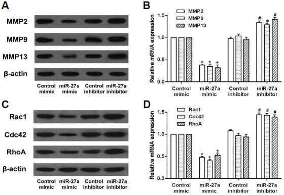
- Fig. 3. Effects of miR-27a on the expression of migration and invasion-related proteins in RA-FLS. RA-FLS were transfected with miR-27a mimic or miR-27a inhibitor. (A) The protein and mRNA expression of MMP2, MMP9, and MMP13 in RA-FLS was detected by western blot and qRT-PCR assay. (B) The protein and mRNA expression of Rac1, Cdc42, and RhoA in RA-FLS was detected by western blot and qRT-PCR assay. * p < 0.05, versus control mimic group. # p < 0.05, versus control inhibitor group.