 Explore
 Validate
 Learn
Learn Immunocytochemistry
Antibody data
- Antibody Data
- Antigen structure
- References [4]
- Comments [0]
- Validations
- Immunocytochemistry [1]
- Immunohistochemistry [2]
- Flow cytometry [1]
Submit
Validation data
Reference
Comment
Report error
- Product number
- PA5-16509 - Provider product page

- Provider
- Invitrogen Antibodies
- Product name
- MMP9 Polyclonal Antibody
- Antibody type
- Polyclonal
- Antigen
- Synthetic peptide
- Description
- PA5-16509 targets MMP-9 (92kDa Collagenase IV) in WB applications and shows reactivity with Guinea Pig and Human samples.
- Concentration
- 1 mg/mL
Submitted references Poria Acid, Triterpenoids Extracted from Poria cocos, Inhibits the Invasion and Metastasis of Gastric Cancer Cells.
Betulonic Acid, as One of the Active Components of the Celastrus orbiculatus Extract, Inhibits the Invasion and Metastasis of Gastric Cancer Cells by Mediating Cytoskeleton Rearrangement In Vitro.
Matrix metalloproteinases 2 and 9 and e-cadherin expression in the endometrium during the implantation window of infertile women before in vitro fertilization treatment.
Effect of the expression of matrix metalloproteases and their tissue inhibitors on survival of patients with resectable colorectal cancer.
Wang H, Luo Y, Chu Z, Ni T, Ou S, Dai X, Zhang X, Liu Y
Molecules (Basel, Switzerland) 2022 Jun 6;27(11)
Molecules (Basel, Switzerland) 2022 Jun 6;27(11)
Betulonic Acid, as One of the Active Components of the Celastrus orbiculatus Extract, Inhibits the Invasion and Metastasis of Gastric Cancer Cells by Mediating Cytoskeleton Rearrangement In Vitro.
Chu Z, Luo Y, Ni T, Zhu M, Feng X, Liu Y, Wang H
Molecules (Basel, Switzerland) 2022 Feb 2;27(3)
Molecules (Basel, Switzerland) 2022 Feb 2;27(3)
Matrix metalloproteinases 2 and 9 and e-cadherin expression in the endometrium during the implantation window of infertile women before in vitro fertilization treatment.
Maia-Filho VO, Rocha AM, Ferreira FP, Bonetti TC, Serafini P, Motta EL
Reproductive sciences (Thousand Oaks, Calif.) 2015 Apr;22(4):416-22
Reproductive sciences (Thousand Oaks, Calif.) 2015 Apr;22(4):416-22
Effect of the expression of matrix metalloproteases and their tissue inhibitors on survival of patients with resectable colorectal cancer.
González L, Eiró N, González LO, Andicoechea A, Barbón E, García-Muñiz JL, Vizoso FJ
Digestive diseases and sciences 2012 Aug;57(8):2063-71
Digestive diseases and sciences 2012 Aug;57(8):2063-71
No comments: Submit comment
Supportive validation
- Submitted by
- Invitrogen Antibodies (provider)
- Main image

- Experimental details
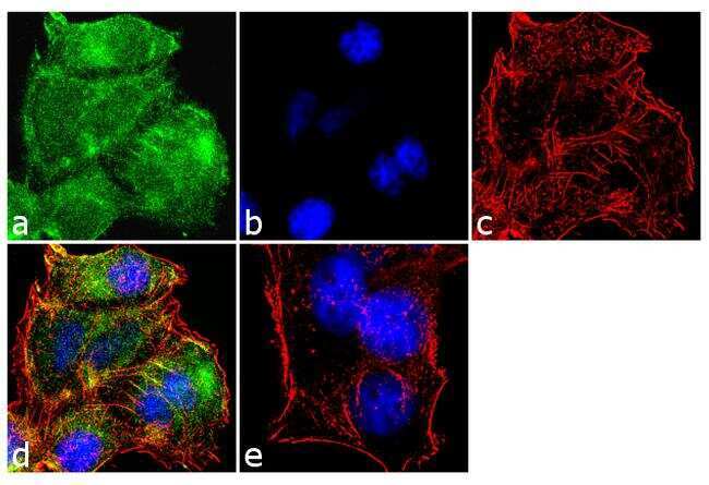
- Immunofluorescence analysis of MMP-9 was performed using 70% confluent log phase MCF-7 cells. The cells were fixed with 4% paraformaldehyde for 10 minutes, permeabilized with 0.1% Triton™ X-100 for 10 minutes, and blocked with 1% BSA for 1 hour at room temperature. The cells were labeled with MMP-9 (Collagenase IV) Rabbit Polyclonal Antibody (Product # PA5-16509) at 2 µg/mL in 0.1% BSA and incubated for 3 hours at room temperature and then labeled with Goat anti-Rabbit IgG (H+L) Superclonal™ Secondary Antibody, Alexa Fluor® 488 conjugate (Product # A27034) at a dilution of 1:2000 for 45 minutes at room temperature (Panel a: green). Nuclei (Panel b: blue) were stained with SlowFade® Gold Antifade Mountant with DAPI (Product # S36938). F-actin (Panel c: red) was stained with Alexa Fluor® 555 Rhodamine Phalloidin (Product # R415, 1:300). Panel d represents the merged image showing cytoplasmic localization. Panel e shows the no primary antibody control. The images were captured at 60X magnification.
Supportive validation
- Submitted by
- Invitrogen Antibodies (provider)
- Main image

- Experimental details
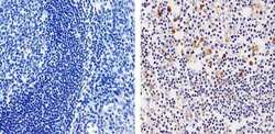
- Immunohistochemistry analysis of MMP-9 (92kDa Collagenase IV) showing staining in the cytoplasm of paraffin-embedded human spleen tissue (right) compared to a negative control without primary antibody (left). To expose target proteins, antigen retrieval was performed using 10mM sodium citrate (pH 6.0), microwaved for 8-15 min. Following antigen retrieval, tissues were blocked in 3% H2O2-methanol for 15 min at room temperature, washed with ddH2O and PBS, and then probed with a MMP-9 (92kDa Collagenase IV) Rabbit Polyclonal Antibody (Product # PA5-16509) diluted in 3% BSA-PBS at a dilution of 1:100 for 1 hour at 37ºC in a humidified chamber. Tissues were washed extensively in PBST and detection was performed using an HRP-conjugated secondary antibody followed by colorimetric detection using a DAB kit. Tissues were counterstained with hematoxylin and dehydrated with ethanol and xylene to prep for mounting.

- Submitted by
- Invitrogen Antibodies (provider)
- Main image
- Experimental details
- Immunohistochemistry analysis of MMP-9 (92kDa Collagenase IV) showing staining in the cytoplasm and weak staining in the nucleus of paraffin-embedded human tonsil tissue (right) compared to a negative control without primary antibody (left). To expose target proteins, antigen retrieval was performed using 10mM sodium citrate (pH 6.0), microwaved for 8-15 min. Following antigen retrieval, tissues were blocked in 3% H2O2-methanol for 15 min at room temperature, washed with ddH2O and PBS, and then probed with a MMP-9 (92kDa Collagenase IV) Rabbit Polyclonal Antibody (Product # PA5-16509) diluted in 3% BSA-PBS at a dilution of 1:20 for 1 hour at 37ºC in a humidified chamber. Tissues were washed extensively in PBST and detection was performed using an HRP-conjugated secondary antibody followed by colorimetric detection using a DAB kit. Tissues were counterstained with hematoxylin and dehydrated with ethanol and xylene to prep for mounting.

Supportive validation
- Submitted by
- Invitrogen Antibodies (provider)
- Main image

- Experimental details
- Flow cytometry analysis of MMP-9 was done on HT-29 cells. Cells were fixed with 70% ethanol for 10 minutes, permeabilized with 0.25% Triton™ X-100 for 20 minutes, and blocked with 5% BSA for 30 minutes at room temperature. Cells were labeled with MMP-9 Rabbit Polyclonal Antibody (PA516509, red histogram) or with rabbit isotype control (pink histogram) at 3-5 ug/million cells in 2.5% BSA. After incubation at room temperature for 2 hours, the cells were labeled with Alexa Fluor® 488 Goat Anti-Rabbit Secondary Antibody (A11008) at a dilution of 1:400 for 30 minutes at room temperature. The representative 10,000 cells were acquired and analyzed for each sample using an Attune® Acoustic Focusing Cytometer. The purple histogram represents unstained control cells and the green histogram represents no-primary-antibody control.
