 Explore
 Validate
 Learn
Learn Western blot
 Immunocytochemistry
Antibody data
- Antibody Data
- Antigen structure
- References [4]
- Comments [0]
- Validations
- Western blot [3]
- Immunohistochemistry [1]
Submit
Validation data
Reference
Comment
Report error
- Product number
- AF2046 - Provider product page

- Provider
- R&D Systems
- Product name
- Human/Mouse GATA-2 Antibody
- Antibody type
- Polyclonal
- Description
- Antigen Affinity-purified. Detects human and mouse GATA-2 in Western blots.
- Reactivity
- Human, Mouse
- Host
- Goat
- Conjugate
- Unconjugated
- Antigen sequence
P23769- Isotype
- IgG
- Vial size
- 100 ug
- Concentration
- LYOPH
- Storage
- Use a manual defrost freezer and avoid repeated freeze-thaw cycles. 12 months from date of receipt, -20 to -70 °C as supplied. 1 month, 2 to 8 °C under sterile conditions after reconstitution. 6 months, -20 to -70 °C under sterile conditions after reconstitution.
Submitted references Piezo1 incorporates mechanical force signals into the genetic program that governs lymphatic valve development and maintenance.
Multiple mouse models of primary lymphedema exhibit distinct defects in lymphovenous valve development.
GATA2 germline mutations impair GATA2 transcription, causing haploinsufficiency: functional analysis of the p.Arg396Gln mutation.
The Down syndrome critical region gene 1 short variant promoters direct vascular bed-specific gene expression during inflammation in mice.
Choi D, Park E, Jung E, Cha B, Lee S, Yu J, Kim PM, Lee S, Hong YJ, Koh CJ, Cho CW, Wu Y, Li Jeon N, Wong AK, Shin L, Kumar SR, Bermejo-Moreno I, Srinivasan RS, Cho IT, Hong YK
JCI insight 2019 Mar 7;4(5)
JCI insight 2019 Mar 7;4(5)
Multiple mouse models of primary lymphedema exhibit distinct defects in lymphovenous valve development.
Geng X, Cha B, Mahamud MR, Lim KC, Silasi-Mansat R, Uddin MKM, Miura N, Xia L, Simon AM, Engel JD, Chen H, Lupu F, Srinivasan RS
Developmental biology 2016 Jan 1;409(1):218-233
Developmental biology 2016 Jan 1;409(1):218-233
GATA2 germline mutations impair GATA2 transcription, causing haploinsufficiency: functional analysis of the p.Arg396Gln mutation.
Cortés-Lavaud X, Landecho MF, Maicas M, Urquiza L, Merino J, Moreno-Miralles I, Odero MD
Journal of immunology (Baltimore, Md. : 1950) 2015 Mar 1;194(5):2190-8
Journal of immunology (Baltimore, Md. : 1950) 2015 Mar 1;194(5):2190-8
The Down syndrome critical region gene 1 short variant promoters direct vascular bed-specific gene expression during inflammation in mice.
Minami T, Yano K, Miura M, Kobayashi M, Suehiro J, Reid PC, Hamakubo T, Ryeom S, Aird WC, Kodama T
The Journal of clinical investigation 2009 Aug;119(8):2257-70
The Journal of clinical investigation 2009 Aug;119(8):2257-70
No comments: Submit comment
Supportive validation
- Submitted by
- R&D Systems (provider)
- Main image

- Experimental details
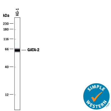
- Detection of Human GATA-2 by Simple WesternTM. Simple Western lane view shows lysates of KG-1 human acute myelogenous leukemia cell line, loaded at 0.2 mg/mL. A specific band was detected for GATA-2 at approximately 64 kDa (as indicated) using 5 µg/mL of Goat Anti-Human GATA-2 Antigen Affinity-purified Polyclonal Antibody (Catalog # AF2046) followed by 1:50 dilution of HRP-conjugated Anti-Goat IgG Secondary Antibody (Catalog # HAF109). This experiment was conducted under reducing conditions and using the 12-230 kDa separation system.
- Submitted by
- R&D Systems (provider)
- Main image

- Experimental details
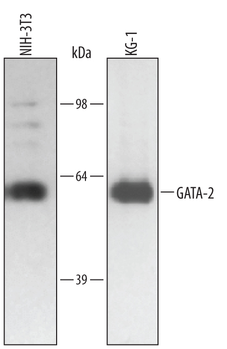
- Detection of Human GATA-2 by Western Blot. Western blot shows lysates of NIH-3T3 mouse embryonic fibroblast cell line and KG-1 human acute myelogenous leukemia cell line. PVDF membrane was probed with 0.5 µg/mL of Human/Mouse GATA-2 Antigen Affinity-purified Polyclonal Antibody (Catalog # AF2046) followed by HRP-conjugated Anti-Goat IgG Secondary Antibody (Catalog # HAF017). A specific band was detected for GATA-2 at approximately 51 kDa (as indicated). This experiment was conducted under reducing conditions and using Immunoblot Buffer Group 1.
- Submitted by
- R&D Systems (provider)
- Main image

- Experimental details
- Detection of Human GATA-2 by Western Blot. Western blot shows lysates of LNCaP human prostate cancer cell line and SH-SY5Y human neuroblastoma cell line. PVDF membrane was probed with 0.5 µg/mL of Goat Anti-Human/Mouse GATA-2 Antigen Affinity-purified Polyclonal Antibody (Catalog # AF2046) followed by HRP-conjugated Anti-Goat IgG Secondary Antibody (Catalog # HAF017). A specific band was detected for GATA-2 at approximately 55 kDa (as indicated). This experiment was conducted under reducing conditions and using Immunoblot Buffer Group 1.

Supportive validation
- Submitted by
- R&D Systems (provider)
- Main image

- Experimental details
- GATA-2 in Human Duodenum. GATA-2 was detected in immersion fixed paraffin-embedded sections of human duodenum (blood vessel) using Goat Anti-Human/Mouse GATA-2 Antigen Affinity-purified Polyclonal Antibody (Catalog # AF2046) at 1 µg/mL for 1 hour at room temperature followed by incubation with the Anti-Goat IgG VisUCyte™ HRP Polymer Antibody (Catalog # VC004). Before incubation with the primary antibody, tissue was subjected to heat-induced epitope retrieval using Antigen Retrieval Reagent-Basic (Catalog # CTS013). Tissue was stained using DAB (brown) and counterstained with hematoxylin (blue). Specific staining was localized to nuclei in endothelial cells. View our protocol for IHC Staining with VisUCyte HRP Polymer Detection Reagents.
