Explore
 Validate
 Learn
Learn Western blot
Antibody data
- Antibody Data
- Antigen structure
- References [3]
- Comments [0]
- Validations
- Western blot [2]
- Flow cytometry [1]
- Other assay [3]
Submit
Validation data
Reference
Comment
Report error
- Product number
- PA5-79532 - Provider product page

- Provider
- Invitrogen Antibodies
- Product name
- CD11b Polyclonal Antibody
- Antibody type
- Polyclonal
- Antigen
- Recombinant full-length protein
- Description
- Reconstitute with 0.2 mL of distilled water to yield a concentration of 500 µg/mL.
- Reactivity
- Human, Mouse, Rat
- Host
- Rabbit
- Isotype
- IgG
- Vial size
- 100 µg
- Concentration
- 500 µg/mL
- Storage
- -20°C
Submitted references Neuroprotective effects of pomegranate (Punica granatum L.) juice and seed extract in paraquat-induced mouse model of Parkinson's disease.
Ceria-based nanotheranostic agent for rheumatoid arthritis.
Sodium para-aminosalicylic acid inhibits manganese-induced NLRP3 inflammasome-dependent pyroptosis by inhibiting NF-κB pathway activation and oxidative stress.
Fathy SM, El-Dash HA, Said NI
BMC complementary medicine and therapies 2021 Apr 26;21(1):130
BMC complementary medicine and therapies 2021 Apr 26;21(1):130
Ceria-based nanotheranostic agent for rheumatoid arthritis.
Kalashnikova I, Chung SJ, Nafiujjaman M, Hill ML, Siziba ME, Contag CH, Kim T
Theranostics 2020;10(26):11863-11880
Theranostics 2020;10(26):11863-11880
Sodium para-aminosalicylic acid inhibits manganese-induced NLRP3 inflammasome-dependent pyroptosis by inhibiting NF-κB pathway activation and oxidative stress.
Peng D, Li J, Deng Y, Zhu X, Zhao L, Zhang Y, Li Z, Ou S, Li S, Jiang Y
Journal of neuroinflammation 2020 Nov 17;17(1):343
Journal of neuroinflammation 2020 Nov 17;17(1):343
No comments: Submit comment
Supportive validation
- Submitted by
- Invitrogen Antibodies (provider)
- Main image

- Experimental details
- Western blot analysis of CD11b in Lane 1: rat spleen tissue lysate, Lane 2: rat thymus tissue lysate, Lane 3: mouse spleen tissue lysate, Lane 4: mouse thymus tissue lysate using 50 µg (reducing conditions) per well. Electrophoresis was performed on 5-20% SDS-PAGE gel at 70V (Stacking gel) / 90V (Resolving gel) for 2-3 hours and protein was transferred to a nitrocellulose membrane at 150mA for 50-90 minutes. Sample was blocked with 5% Non-fat Milk/TBS for 1.5 hours at room temperature, incubated with CD11b polyclonal antibody (Product # PA5-79532) at a dilution of 0.5 µg/mL (overnight at 4°C), followed by goat anti-rabbit IgG-HRP secondary antibody at a dilution of 1:10,000. Signal development was performed using a chemiluminescence (ECL) kit.

- Submitted by
- Invitrogen Antibodies (provider)
- Main image

- Experimental details
- Western blot was performed using Anti-CD11b Polyclonal Antibody (Product # PA5-79532) and a 165 kDa band corresponding to CD11b was observed across cell lines tested except in U937, HL-60, Jurkat and HeLa. Whole cell extracts (30 µg lysate) of U-937 (Lane 1), U-937 treated with 10ng/mL TPA for 3 days (Lane 2), HL-60 (Lane 3), HL-60 treated with 1µM RA for 6 days (Lane 4), THP1 (Lane 5), THP1 treated with 10ng/mL TPA for 6 days (Lane 6), Jurkat (Lane 7) and HeLa (Lane 8) were electrophoresed using Novex® NuPAGE® 4-12 % Bis-Tris gel (Product # NP0321BOX). Resolved proteins were then transferred onto a nitrocellulose membrane (Product # IB23001) by iBlot® 2 Dry Blotting System (Product # IB21001). The blot was probed with the primary antibody (1:1000 dilution) and detected by chemiluminescence with Goat anti-Rabbit IgG (H+L), Superclonal™ Recombinant Secondary Antibody, HRP (Product # A27036, 1:4000 dilution) using the iBright FL 1000 (Product # A32752). Chemiluminescent detection was performed using Novex® ECL Chemiluminescent Substrate. An uncharacterized band (*) around 35 kDa was seen across cell lines tested.

Supportive validation
- Submitted by
- Invitrogen Antibodies (provider)
- Main image
- Experimental details
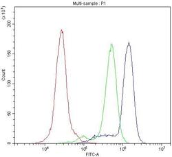
- Flow Cytometry of CD11b in HL-60 cells (blue line), isotype control rabbit IgG (green line) and unlabeled (red line). Samples were blocked with 10% goat serum, incubated with CD11b Polyclonal Antibody (Product # PA5-79532) at a dilution of 1 μg (per 1x10^6 cells), followed by DyLight®488 conjugated goat anti-rabbit IgG (for 30 minutes at 20°C) using 5-10 μg (per 1x10^6 cells) dilution.
Supportive validation
- Submitted by
- Invitrogen Antibodies (provider)
- Main image
- Experimental details
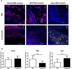
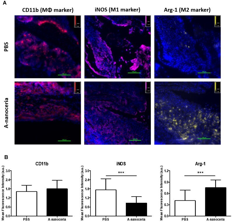
- Figure 7 Macrophage polarization in tissue sections harvested from PBS and A-nanoceria treated groups of CIA mice by immunohistology. (A) Images of tissue sections from animals treated with PBS or A-nanoceria. Sections of inflamed tissues (DAPI in blue) displayed a high concentration of infiltrating macrophages into the paws (CD11b in red). The number of M1 macrophages (iNOS in pink) decreased upon treatment with A-nanoceria. Tissues from animals treated with A-nanoceria revealed a high number of M2 macrophages (Arg-1 in yellow). Immunohistology of tissue sections harvested from normal mice can be found in Figure S6 ; (B) Summarized plot of CD11b, iNOS and Arg-1 MFI signals in ROI from images. In each group, we took 5 fluorescence images from two mice tissues and plotted 5 random ROIs for each image (N = 25. error bar = SD, p -value: *** p < 0.0005).
- Submitted by
- Invitrogen Antibodies (provider)
- Main image
- Experimental details
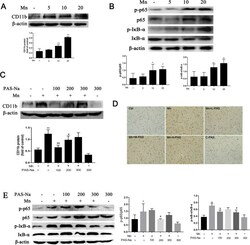
- Fig. 4 PAS-Na mitigates Mn-induced BV2 cells activation and microglia proliferation in basal ganglia of rats by inhibited NF-kappaB pathway activation. a , b The rats were treated with 5, 10, and 20 mg/kg MnCl 2 for 8 weeks. a The protein expressions of CD11b in the basal ganglia of rats was analyzed by western blot ( n = 4 per group). b The protein expressions of p-p65, p65, p-IkappaB-alpha, and IkappaB-alpha in the basal ganglia of rats was detected by western blot ( n = 4 per group). c - d The rats were treated with 20 mg/kg MnCl 2 for 8 weeks and then treated with 100, 200, and 300 mg/kg PAS-Na for an additional 6 weeks. c The protein expression of CD11b in the basal ganglia of rats were analyzed by western blot ( n = 4 per group). d Immunohistochemical results of CD11b in the basal ganglia of rats ( n = 3 per group). Scale bars: 100 mum. e The protein expressions of p-p65, p65, p-IkappaB-alpha, and IkappaB-alpha in the basal ganglia of rats were detected by western blot ( n = 4 per group). The protein expression was normalized by beta-actin or corresponding total protein content. Data are presented as mean +- SD. * p < 0.05 and ** p < 0.01: significant as compared to the control group; # p < 0.05 and ## p < 0.01: significant as compared to corresponding Mn-treated group

- Submitted by
- Invitrogen Antibodies (provider)
- Main image

- Experimental details
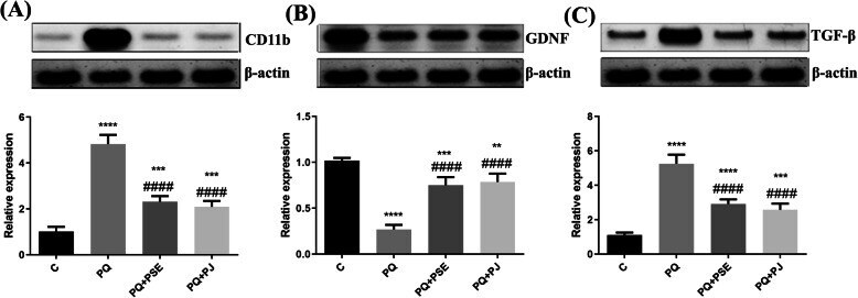
- Fig. 7 Representative cropped blots with relative expression levels of the striatal CD11b, TGF-beta, and GDNF in different animal groups. The full length blots are presented in supplementary figure 3 . Relative expression of CD11b ( a ), GDNF ( b ), and TGF-beta ( c ) in striatum. C = control group; PQ = Paraquat (alone)-induced group; PQ+PSE = PQ-induced group treated with PSE; PQ+PJ = PQ-induced group treated with PJ. Data are expressed as mean +- S.D. of 10 mice in each group. **: P < 0.01, ***: P < 0.001, and ****: P < 0.0001 compared with control group; ####: P < 0.0001 compared with PQ group