Explore
 Validate
 Learn
Learn Western blot
Antibody data
- Antibody Data
- Antigen structure
- References [3]
- Comments [0]
- Validations
- Western blot [3]
- Immunocytochemistry [3]
- Immunohistochemistry [3]
- Other assay [1]
Submit
Validation data
Reference
Comment
Report error
- Product number
- PA5-90724 - Provider product page

- Provider
- Invitrogen Antibodies
- Product name
- CD11b Polyclonal Antibody
- Antibody type
- Polyclonal
- Antigen
- Recombinant full-length protein
- Description
- Immunogen sequence: PQEDSDIAFL IDGSGSIIPH DFRRMKEFVS TVMEQLKKSK TLFSLMQYSE EFRIHFTFKE FQNNPNPRSL VKPITQLLGR THTATGIRKV VRELFNITNG ARKNAFKILV VITDGEKFGD PLGYEDVIPE ADREGVIRYV IGVGDAFRSE KSRQELNTIA SKPPRDHVFQ VNNFEALKTI QNQLREKIFA IEGTQT
- Concentration
- 1.64 mg/mL
Submitted references Blending with transition metals improves bioresorbable zinc as better medical implants.
Targeting myeloid-derived suppressor cells to attenuate vasculogenic mimicry and synergistically enhance the anti-tumor effect of PD-1 inhibitor.
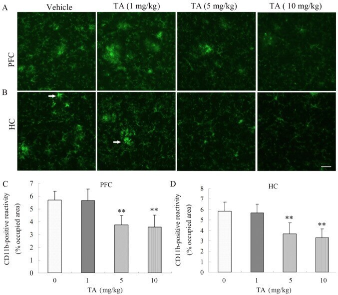
Neuroprotective effect of tormentic acid against memory impairment and neuro‑inflammation in an Alzheimer's disease mouse model.
Su Y, Fu J, Zhou J, Georgas E, Du S, Qin YX, Wang Y, Zheng Y, Zhu D
Bioactive materials 2023 Feb;20:243-258
Bioactive materials 2023 Feb;20:243-258
Targeting myeloid-derived suppressor cells to attenuate vasculogenic mimicry and synergistically enhance the anti-tumor effect of PD-1 inhibitor.
Li Y, Qiao K, Zhang X, Liu H, Zhang H, Li Z, Liu Y, Sun T
iScience 2021 Dec 17;24(12):103392
iScience 2021 Dec 17;24(12):103392
Neuroprotective effect of tormentic acid against memory impairment and neuro‑inflammation in an Alzheimer's disease mouse model.
Cui W, Sun C, Ma Y, Wang S, Wang X, Zhang Y
Molecular medicine reports 2020 Aug;22(2):739-750
Molecular medicine reports 2020 Aug;22(2):739-750
No comments: Submit comment
Supportive validation
- Submitted by
- Invitrogen Antibodies (provider)
- Main image

- Experimental details
- Western blot analysis of extracts of various cell lines, using ITGAM Polyclonal antibody (Product # PA5-90724) at 1:1000 dilution. Secondary antibody: HRP Goat Anti-Rabbit IgG (H+L) at 1:10000 dilution. Lysates/proteins: 25ug per lane. Blocking buffer: 3% nonfat dry milk in TBST.

- Submitted by
- Invitrogen Antibodies (provider)
- Main image
- Experimental details
- Western Blot analysis of CD11b in extracts of various cell lines using CD11b Polyclonal Antibody (Product # PA5-90724) at a dilution of 1:1000. A HRP Goat Anti-Rabbit IgG (H+L) secondary antibody was used at a dilution of 1:10,000. Lysates/proteins: 25 µg per lane. Blocking buffer: 3% nonfat dry milk in TBST.

- Submitted by
- Invitrogen Antibodies (provider)
- Main image

- Experimental details
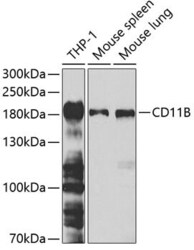
- Western blot was performed using Anti-CD11b Polyclonal Antibody (Product # PA5-90724) and a 165 kDa band corresponding to CD11b was detected across THP-1, mouse spleen, and rat spleen and not detected in Jurkat and mouse heart. Uncharacterized bands (*) were also detected at 80-90 kDa. Whole-cell extracts (30 µg lysate) of THP1 (Lane 1) and Jurkat (Lane 2), mouse spleen (Lane 3), mouse heart (Lane 4), and Rat spleen (Lane 5) were electrophoresed using NuPAGE™ 4-12% Bis-Tris Protein Gel (Product # NP0321BOX), 10 well. Resolved proteins were then transferred onto a nitrocellulose membrane (Product # IB23001) by iBlot® 2 Dry Blotting System (Product # IB21001). The blot was probed with the primary antibody (1:1000 dilution) and detected by chemiluminescence with Goat anti-Rabbit IgG (H+L) Superclonal™ Recombinant Secondary Antibody, HRP (Product # A27036, 1:20000 dilution) using the iBright™ FL1500 Imaging System (Product # A44115). Chemiluminescent detection was performed using SuperSignal™ West Pico PLUS Chemiluminescent Substrate (Product # 34580).

Supportive validation
- Submitted by
- Invitrogen Antibodies (provider)
- Main image
- Experimental details
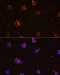
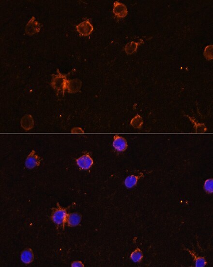
- Immunocytochemistry-Immunofluorescence analysis of CD11b was performed in THP-1 cells using CD11b Polyclonal Antibody (Product # PA5-90724).
- Submitted by
- Invitrogen Antibodies (provider)
- Main image
- Experimental details
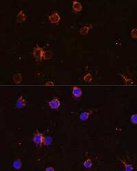
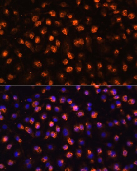
- Immunocytochemistry-Immunofluorescence analysis of CD11b was performed in RAW264.7 cells using CD11b Polyclonal Antibody (Product # PA5-90724) at a dilution of 1:100. Blue: DAPI for nuclear staining.
- Submitted by
- Invitrogen Antibodies (provider)
- Main image
- Experimental details
- Immunocytochemistry-Immunofluorescence analysis of CD11b was performed in THP-1 cells using CD11b Polyclonal Antibody (Product # PA5-90724).
Supportive validation
- Submitted by
- Invitrogen Antibodies (provider)
- Main image

- Experimental details
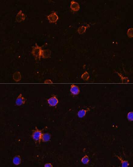
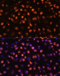
- Immunohistochemistry-Immunofluorescence analysis of CD11b was performed in rat bone marrow tissue using CD11b Polyclonal Antibody (Product # PA5-90724) at a dilution of 1:100. Blue: DAPI for nuclear staining.

- Submitted by
- Invitrogen Antibodies (provider)
- Main image

- Experimental details
- Immunohistochemical analysis of CD11b was performed using formalin-fixed paraffin-embedded mouse spleen tissue sections. To expose the target protein, heat-induced epitope retrieval was performed on de-paraffinized sections using eBioscience™ IHC Antigen Retrieval Solution - High pH (10X) (Product # 00-4956-58) diluted to 1X solution in water in a decloaking chamber at 110 degree Celsius for 15 minutes. Following antigen retrieval, the sections were blocked with 2% normal goat serum in 1X PBS for 45 minutes at room temperature and then probed with or without CD11b Polyclonal Antibody (Product # PA5-90724) at 1:100 dilution in 0.1% normal goat serum overnight at 4 degree Celsius in a humidified chamber. Detection was performed using Goat anti-Rabbit IgG (H+L) Highly Cross-Adsorbed Secondary Antibody, Alexa Fluor Plus 488 (Product # A32731) at a dilution of 1:2000 in 0.1% normal goat serum for 45 minutes at room temperature. Nuclei were stained with DAPI (Product # D1306) and the sections were mounted using ProLong™ Glass Antifade Mountant (Product # P36984). The images were captured on EVOS™ M7000 Imaging System (Product # AMF7000) at 20X magnification.

Supportive validation
- Submitted by
- Invitrogen Antibodies (provider)
- Main image
- Experimental details
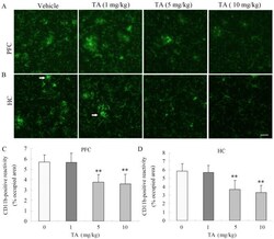
- Figure 3. TA treatment decreases the area of CD11b-positive cells in amyloid beta precursor protein/presenilin 1 transgenic mice. Cells were visualized by immunohistochemical staining and the area of CD11b-positive cells was compared with the vehicle treatment group. (A) CD11b-positive cells in the PFC, indicated by white arrow. (B) CD11b-positive cells in the HC. Scale bar, 40 um. (C) Area of CD11b-positive cells in the PFC. (D) Area of CD11b-positive cells in the HC. Data are expressed as the mean +- SEM for the immunopositive area as % of the total surface area. n=5 in each group. **P