Explore
 Validate
 Learn
Learn Flow cytometry
Antibody data
- Antibody Data
- Antigen structure
- References [4]
- Comments [0]
- Validations
- Flow cytometry [1]
- Other assay [1]
Submit
Validation data
Reference
Comment
Report error
- Product number
- 50-7823-42 - Provider product page

- Provider
- Invitrogen Antibodies
- Product name
- IL-23 p19 Monoclonal Antibody (23dcdp), eFluor™ 660, eBioscience™
- Antibody type
- Monoclonal
- Antigen
- Other
- Description
- Description: This 23dcdp monoclonal antibody reacts with the p19 subunit of human IL-23. This heterodimeric cytokine is composed of two disulfide-linked subunits, p40 and p19. It is closely related to IL-12, with which it shares the p40 subunit. The IL-23 receptor is also heterodimeric and shares the IL-12Rbeta1 chain with IL-12, while the IL-23R chain is unique to the IL-23 receptor complex. IL-23R signaling occurs through the Jak/STAT pathway and results in RORgammat expression, which promotes maintenance and proliferation of T helper 17 (Th17) cells.
- Antibody clone number
- 23dcdp
- Concentration
- 5 µL/Test
Submitted references Lack of Mucosal Cholinergic Innervation Is Associated With Increased Risk of Enterocolitis in Hirschsprung's Disease.
Antibiotic-induced microbiome perturbations are associated with significant alterations to colonic mucosal immunity in rhesus macaques.
Prostaglandin E2 inhibits IL-23 and IL-12 production by human monocytes through down-regulation of their common p40 subunit.
Abundant expression of the interleukin (IL)23 subunit p19, but low levels of bioactive IL23 in the rheumatoid synovium: differential expression and Toll-like receptor-(TLR) dependent regulation of the IL23 subunits, p19 and p40, in rheumatoid arthritis.
Keck S, Galati-Fournier V, Kym U, Moesch M, Usemann J, Müller I, Subotic U, Tharakan SJ, Krebs T, Stathopoulos E, Schmittenbecher P, Cholewa D, Romero P, Reingruber B, Bruder E, Group NS, Holland-Cunz S
Cellular and molecular gastroenterology and hepatology 2021;12(2):507-545
Cellular and molecular gastroenterology and hepatology 2021;12(2):507-545
Antibiotic-induced microbiome perturbations are associated with significant alterations to colonic mucosal immunity in rhesus macaques.
Manuzak JA, Zevin AS, Cheu R, Richardson B, Modesitt J, Hensley-McBain T, Miller C, Gustin AT, Coronado E, Gott T, Fang M, Cartwright M, Wangari S, Agricola B, May D, Smith E, Hampel HB, Gale M, Cameron CM, Cameron MJ, Smedley J, Klatt NR
Mucosal immunology 2020 May;13(3):471-480
Mucosal immunology 2020 May;13(3):471-480
Prostaglandin E2 inhibits IL-23 and IL-12 production by human monocytes through down-regulation of their common p40 subunit.
Kalim KW, Groettrup M
Molecular immunology 2013 Mar;53(3):274-82
Molecular immunology 2013 Mar;53(3):274-82
Abundant expression of the interleukin (IL)23 subunit p19, but low levels of bioactive IL23 in the rheumatoid synovium: differential expression and Toll-like receptor-(TLR) dependent regulation of the IL23 subunits, p19 and p40, in rheumatoid arthritis.
Brentano F, Ospelt C, Stanczyk J, Gay RE, Gay S, Kyburz D
Annals of the rheumatic diseases 2009 Jan;68(1):143-50
Annals of the rheumatic diseases 2009 Jan;68(1):143-50
No comments: Submit comment
Supportive validation
- Submitted by
- Invitrogen Antibodies (provider)
- Main image
- Experimental details
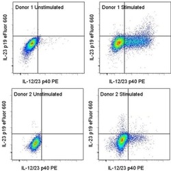
- Intracellular staining of unstimulated (left) or LPS-stimulated and monensin-treated (right), in vitro-cultured human dendritic cells with Anti-Human IL-12/23 p40 PE (Product # 12-7235-42) and Anti-Human IL-23 p19 eFluor® 660. Cultures were fixed and permeabilized with the Fixation and Permeabilization Kit (Product # 88-8823-88). Total viable cells as determined by staining with the Fixable Viability Dye eFluor® 450 (Product # 65-0863-14) were used for analysis. Data from two donors are shown to illustrate variability in expression.

Supportive validation
- Submitted by
- Invitrogen Antibodies (provider)
- Main image

- Experimental details
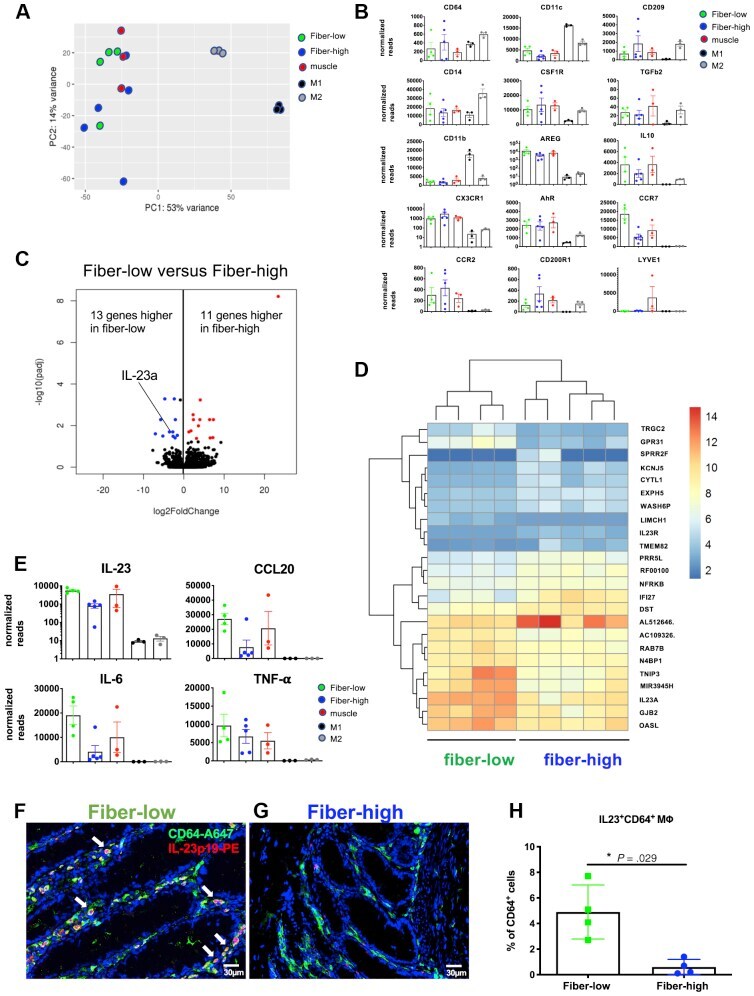
- Figure 11 MPhi isolated from fiber-high colonic tissue express diminished IL23 levels. RNA sequencing was performed on sorted viable colonic MPhi and blood-derived M1 and M2 MPhi. ( A ) PCA plot shows similarity between the different populations based on their first 2 principal components. The top 500 genes, selected by highest row variance, were used. Fiber-low ( green , n = 4); fiber-high ( blue , n = 5); muscle ( red , n = 3); M1 ( black , n = 3); M2 ( gray , n = 3). ( B ) Normalized sequence reads of selected macrophage-related genes. Scatter plots with bar show means +- standard error of the mean. ( C ) Global transcriptional changes between macrophages isolated from fiber-low colonic tissue and fiber-high colonic tissue visualized by a volcano plot. Comparison of gene expression between the groups of samples was performed with the package DESeq2. The Wald test was used to generate P values and log2 fold changes. Genes with an adjusted P value