 Explore
 Validate
 Learn
Learn Western blot
Antibody data
- Antibody Data
- Antigen structure
- References [4]
- Comments [0]
- Validations
- Western blot [4]
- Immunohistochemistry [1]
- Flow cytometry [1]
- Other assay [4]
Submit
Validation data
Reference
Comment
Report error
- Product number
- MA1-46412 - Provider product page

- Provider
- Invitrogen Antibodies
- Product name
- HAP1 Monoclonal Antibody (1B6)
- Antibody type
- Monoclonal
- Antigen
- Other
- Description
- In Western blot bands can be seen ~95 kDa for isoform A and ~110 kDa for isoform B.
- Antibody clone number
- 1B6
- Concentration
- 1 mg/mL
Submitted references Sequential dynein effectors regulate axonal autophagosome motility in a maturation-dependent pathway.
Mutant huntingtin impairs PNKP and ATXN3, disrupting DNA repair and transcription.
Huntingtin-associated protein 1: Eutherian adaptation from a TRAK-like protein, conserved gene promoter elements, and localization in the human intestine.
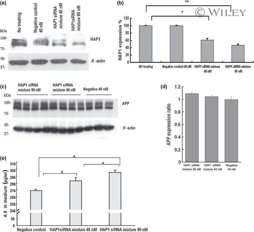
Huntingtin associated protein 1 regulates trafficking of the amyloid precursor protein and modulates amyloid beta levels in neurons.
Cason SE, Carman PJ, Van Duyne C, Goldsmith J, Dominguez R, Holzbaur ELF
The Journal of cell biology 2021 Jul 5;220(7)
The Journal of cell biology 2021 Jul 5;220(7)
Mutant huntingtin impairs PNKP and ATXN3, disrupting DNA repair and transcription.
Gao R, Chakraborty A, Geater C, Pradhan S, Gordon KL, Snowden J, Yuan S, Dickey AS, Choudhary S, Ashizawa T, Ellerby LM, La Spada AR, Thompson LM, Hazra TK, Sarkar PS
eLife 2019 Apr 17;8
eLife 2019 Apr 17;8
Huntingtin-associated protein 1: Eutherian adaptation from a TRAK-like protein, conserved gene promoter elements, and localization in the human intestine.
Lumsden AL, Young RL, Pezos N, Keating DJ
BMC evolutionary biology 2016 Oct 13;16(1):214
BMC evolutionary biology 2016 Oct 13;16(1):214
Huntingtin associated protein 1 regulates trafficking of the amyloid precursor protein and modulates amyloid beta levels in neurons.
Yang GZ, Yang M, Lim Y, Lu JJ, Wang TH, Qi JG, Zhong JH, Zhou XF
Journal of neurochemistry 2012 Sep;122(5):1010-22
Journal of neurochemistry 2012 Sep;122(5):1010-22
No comments: Submit comment
Supportive validation
- Submitted by
- Invitrogen Antibodies (provider)
- Main image

- Experimental details
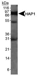
- Detection of HAP1 in mouse brain lysate using Product # MA1-46412.
- Submitted by
- Invitrogen Antibodies (provider)
- Main image
- Experimental details
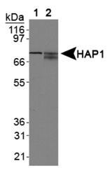
- Western blot analysis of HAP1 using Lane 1: mouse brain lysate and Lane 2: human brain lysate.

- Submitted by
- Invitrogen Antibodies (provider)
- Main image

- Experimental details
- Western blot analysis of HAP1 in Lane 1: mouse brain lysate and Lane 2: human brain lysate. Samples were incubated in HAP1 monoclonal antibody (Product # MA1-46412).

- Submitted by
- Invitrogen Antibodies (provider)
- Main image

- Experimental details
- Western blot analysis of HAP1 in mouse brain lysate. Sample was incubated in HAP1 monoclonal antibody (Product # MA1-46412).

Supportive validation
- Submitted by
- Invitrogen Antibodies (provider)
- Main image
- Experimental details
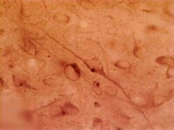
- Immunohistochemical analysis of HAP1 in rat hypothalamus. Samples were incubated in HAP1 monoclonal antibody (Product # MA1-46412).

Supportive validation
- Submitted by
- Invitrogen Antibodies (provider)
- Main image

- Experimental details
- Flow cytometry of HAP1 in 1 x 10^6 MCF-7 cells. Samples were incubated in HAP1 monoclonal antibody (Product # MA1-46412) using a dilution of 1 µg/1x10^6 cells. Antibody (dark blue). Isotype control shown in orange.

Supportive validation
- Submitted by
- Invitrogen Antibodies (provider)
- Main image

- Experimental details
- NULL
- Submitted by
- Invitrogen Antibodies (provider)
- Main image

- Experimental details
- NULL

- Submitted by
- Invitrogen Antibodies (provider)
- Main image
- Experimental details
- NULL
- Submitted by
- Invitrogen Antibodies (provider)
- Main image

- Experimental details
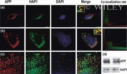
- Fig. 5 HAP1 and 5-HT localization in villi from healthy human duodenum. ( a-e ) are related images highlighting an example of cells individually positive for HAP1 or 5-HT. Images ( a-d ) are the same field of view (enlarged from e ), showing HAP1 alone ( a ), 5-HT alone ( b ), merged ( c ), merged with DAPI nuclei stain ( d ), with lumen to the left. Image e shows HAP1, 5-HT and DAPI staining in the wider context of the villus. Images ( f-j ) highlight a cell immunopositive for both HAP1 and 5-HT. Lumen is to the top right in ( f-i ). Epifluorescence images taken with 40x magnification. Scale bar = 20 mum
