 Explore
 Validate
 Learn
LearnMAB7045
antibody from Novus Biologicals
Targeting: CD44
CD44R, CSPG8, HCELL, IN, MC56, MDU2, MDU3, MIC4, Pgp1
 Western blot
 Immunocytochemistry
Antibody data
- Antibody Data
- Antigen structure
- References [2]
- Comments [0]
- Validations
- Western blot [1]
- Immunohistochemistry [1]
- Flow cytometry [1]
Submit
Validation data
Reference
Comment
Report error
- Product number
- MAB7045 - Provider product page

- Provider
- Novus Biologicals
- Product name
- Mouse Monoclonal CD44 Antibody
- Antibody type
- Monoclonal
- Description
- Protein A or G purified from hybridoma culture supernatant. Detects human CD44 in direct ELISAs and Western blots. In direct ELISAs, no cross-reactivity with recombinant CD44 from mouse, rat, or pig is observed.
- Reactivity
- Human
- Host
- Mouse
- Isotype
- IgG
- Vial size
- 100 ug
- Concentration
- LYOPH
- Storage
- Use a manual defrost freezer and avoid repeated freeze-thaw cycles. 12 months from date of receipt, -20 to -70 degreesC as supplied. 1 month, 2 to 8 degreesC under sterile conditions after reconstitution. 6 months, -20 to -70 degreesC under sterile conditions after reconstitution.
Submitted references Clinical impact of different exosomes' protein expression in pancreatic ductal carcinoma patients treated with standard first line palliative chemotherapy.
SALL4 promotes gastric cancer progression through activating CD44 expression.
Giampieri R, Piva F, Occhipinti G, Bittoni A, Righetti A, Pagliaretta S, Murrone A, Bianchi F, Amantini C, Giulietti M, Ricci G, Principato G, Santoni G, Berardi R, Cascinu S
PloS one 2019;14(5):e0215990
PloS one 2019;14(5):e0215990
SALL4 promotes gastric cancer progression through activating CD44 expression.
Yuan X, Zhang X, Zhang W, Liang W, Zhang P, Shi H, Zhang B, Shao M, Yan Y, Qian H, Xu W
Oncogenesis 2016 Nov 7;5(11):e268
Oncogenesis 2016 Nov 7;5(11):e268
No comments: Submit comment
Supportive validation
- Submitted by
- Novus Biologicals (provider)
- Main image
- Experimental details
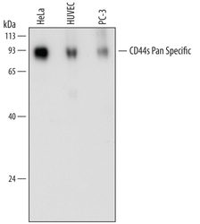
- Detection of Human CD44 by Western Blot. Western blot shows lysates of HeLa human cervical epithelial carcinoma cell line, HUVEC human umbilical vein endothelial cells, and PC-3 human prostate cancer cell line. PVDF membrane was probed with 0.2 µg/mL of Mouse Anti-Human CD44s Pan Specific Monoclonal Antibody (Catalog # MAB7045) followed by HRP-conjugated Anti-Mouse IgG Secondary Antibody (Catalog # HAF007). Specific bands were detected for CD44 at approximately 80 to 100 kDa (as indicated). This experiment was conducted under reducing conditions and using Immunoblot Buffer Group 1.

Supportive validation
- Submitted by
- Novus Biologicals (provider)
- Main image
- Experimental details
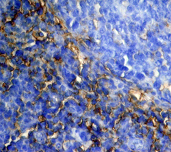
- CD44 in Human Tonsil. CD44 was detected in immersion fixed paraffin-embedded sections of human tonsil using Mouse Anti-Human CD44s Pan Specific Monoclonal Antibody (Catalog # MAB7045) at 15 µg/mL overnight at 4 °C. Tissue was stained using the Anti-Mouse HRP-DAB Cell & Tissue Staining Kit (brown; Catalog # CTS002) and counterstained with hematoxylin (blue). View our protocol for Chromogenic IHC Staining of Paraffin-embedded Tissue Sections.

Supportive validation
- Submitted by
- Novus Biologicals (provider)
- Main image
- Experimental details
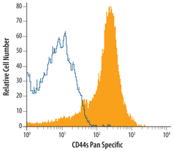
- Detection of CD44 in Human Blood Lymphocytes by Flow Cytometry. Human peripheral blood lymphocytes were stained with Mouse Anti-Human CD44s Pan Specific Monoclonal Antibody (Catalog # MAB7045, filled histogram) or isotype control antibody (Catalog # MAB003, open histogram), followed by Allophy-cocyanin-conjugated Anti-Mouse IgG Secondary Antibody (Catalog # F0101B).
