 Explore
 Validate
 Learn
LearnMA4405
antibody from Invitrogen Antibodies
Targeting: CD44
CD44R, CSPG8, HCELL, IN, MC56, MDU2, MDU3, MIC4, Pgp1
 Western blot
 Immunocytochemistry
Antibody data
- Antibody Data
- Antigen structure
- References [11]
- Comments [0]
- Validations
- Immunocytochemistry [3]
- Flow cytometry [2]
Submit
Validation data
Reference
Comment
Report error
- Product number
- MA4405 - Provider product page

- Provider
- Invitrogen Antibodies
- Product name
- CD44 Monoclonal Antibody (1M7.8.1)
- Antibody type
- Monoclonal
- Antigen
- Other
- Description
- MA4405 targets CD44 in FACS, IA, IF, IP, and WB applications and shows reactivity with mouse and Human samples. The MA4405 immunogen is mouse 80-95 kD lymphocyte surface glycoprotein H-CAM (CD44). MA4405 detects a standard 85-kDa isoform of CD44 and a number of high molecular mass variant isoforms. This antibody is produced by injecting Rat IgG secreting hybridoma cells into the peritoneum of mice. The resulting ascites is collected from the mice and the antibody is purified. This product is a Low Endotoxin formulation. This product has been tested for endotoxins by limulus amoebocyte lysate (LAL) assay and contains an endotoxin concentration of less than or equal to 10 endotoxin units per milligram (EU/mg).
- Reactivity
- Human, Mouse
- Host
- Rat
- Isotype
- IgG
- Antibody clone number
- 1M7.8.1
- Vial size
- 500 µg
- Concentration
- 1 mg/mL
- Storage
- -20°C
Submitted references Enrichment of retinal ganglion and Müller glia progenitors from retinal organoids derived from human induced pluripotent stem cells - possibilities and current limitations.
Hyaluronic acid, CD44 and RHAMM regulate myoblast behavior during embryogenesis.
Exploiting Arginine Auxotrophy with Pegylated Arginine Deiminase (ADI-PEG20) to Sensitize Pancreatic Cancer to Radiotherapy via Metabolic Dysregulation.
DOCK8 deficiency impairs CD8 T cell survival and function in humans and mice.
Lymphoid environment limits superantigen and antigen-induced T cell proliferation at high precursor frequency.
Analysis of CD44-containing lipid rafts: Recruitment of annexin II and stabilization by the actin cytoskeleton.
Resistance to murine AIDS in offspring of mice infected with LP-BM5. Role of CD8 T cells.
Intervention of CD4+ cell subset shifts and autoimmunity in the BXSB mouse by murine CTLA4Ig.
Distinct phenotypes of antigen-selected CD8 T cells emerge at different stages of an in vivo immune response.
Genetic characterization of a polymorphic murine cell-surface glycoprotein.
Genetic characterization of a polymorphic murine cell-surface glycoprotein.
Freude KK, Saruhanian S, McCauley A, Paterson C, Odette M, Oostenink A, Hyttel P, Gillies M, Haukedal H, Kolko M
World journal of stem cells 2020 Oct 26;12(10):1171-1183
World journal of stem cells 2020 Oct 26;12(10):1171-1183
Hyaluronic acid, CD44 and RHAMM regulate myoblast behavior during embryogenesis.
Leng Y, Abdullah A, Wendt MK, Calve S
Matrix biology : journal of the International Society for Matrix Biology 2019 May;78-79:236-254
Matrix biology : journal of the International Society for Matrix Biology 2019 May;78-79:236-254
Exploiting Arginine Auxotrophy with Pegylated Arginine Deiminase (ADI-PEG20) to Sensitize Pancreatic Cancer to Radiotherapy via Metabolic Dysregulation.
Singh PK, Deorukhkar AA, Venkatesulu BP, Li X, Tailor R, Bomalaski JS, Krishnan S
Molecular cancer therapeutics 2019 Dec;18(12):2381-2393
Molecular cancer therapeutics 2019 Dec;18(12):2381-2393
DOCK8 deficiency impairs CD8 T cell survival and function in humans and mice.
Randall KL, Chan SS, Ma CS, Fung I, Mei Y, Yabas M, Tan A, Arkwright PD, Al Suwairi W, Lugo Reyes SO, Yamazaki-Nakashimada MA, Garcia-Cruz Mde L, Smart JM, Picard C, Okada S, Jouanguy E, Casanova JL, Lambe T, Cornall RJ, Russell S, Oliaro J, Tangye SG, Bertram EM, Goodnow CC
The Journal of experimental medicine 2011 Oct 24;208(11):2305-20
The Journal of experimental medicine 2011 Oct 24;208(11):2305-20
Lymphoid environment limits superantigen and antigen-induced T cell proliferation at high precursor frequency.
Attinger A, MacDonald HR, Acha-Orbea H
European journal of immunology 2001 Mar;31(3):884-93
European journal of immunology 2001 Mar;31(3):884-93
Analysis of CD44-containing lipid rafts: Recruitment of annexin II and stabilization by the actin cytoskeleton.
Oliferenko S, Paiha K, Harder T, Gerke V, Schwärzler C, Schwarz H, Beug H, Günthert U, Huber LA
The Journal of cell biology 1999 Aug 23;146(4):843-54
The Journal of cell biology 1999 Aug 23;146(4):843-54
Resistance to murine AIDS in offspring of mice infected with LP-BM5. Role of CD8 T cells.
Pavlovitch JH, Hulier E, Rizk-Rabin M, Marussig M, Mazier D, Joffret ML, Hoos S, Papiernik M
Journal of immunology (Baltimore, Md. : 1950) 1996 Jun 15;156(12):4757-63
Journal of immunology (Baltimore, Md. : 1950) 1996 Jun 15;156(12):4757-63
Intervention of CD4+ cell subset shifts and autoimmunity in the BXSB mouse by murine CTLA4Ig.
Chu EB, Hobbs MV, Wilson CB, Romball CG, Linsley PS, Weigle WO
Journal of immunology (Baltimore, Md. : 1950) 1996 Feb 1;156(3):1262-8
Journal of immunology (Baltimore, Md. : 1950) 1996 Feb 1;156(3):1262-8
Distinct phenotypes of antigen-selected CD8 T cells emerge at different stages of an in vivo immune response.
Walker PR, Ohteki T, Lopez JA, MacDonald HR, Maryanski JL
Journal of immunology (Baltimore, Md. : 1950) 1995 Oct 1;155(7):3443-52
Journal of immunology (Baltimore, Md. : 1950) 1995 Oct 1;155(7):3443-52
Genetic characterization of a polymorphic murine cell-surface glycoprotein.
Lesley J, Trowbridge IS
Immunogenetics 1982 Mar;15(3):313-20
Immunogenetics 1982 Mar;15(3):313-20
Genetic characterization of a polymorphic murine cell-surface glycoprotein.
Lesley J, Trowbridge IS
Immunogenetics 1982 Mar;15(3):313-20
Immunogenetics 1982 Mar;15(3):313-20
No comments: Submit comment
Supportive validation
- Submitted by
- Invitrogen Antibodies (provider)
- Main image
- Experimental details
- Immunofluorescent analysis of CD44 (green) showing staining in the membrane of BAF-3 cells (right) compared to a negative control without primary antibody (left). Formalin-fixed cells were permeabilized with 0.1% Triton X-100 in TBS for 5-10 minutes and blocked with 3% BSA-PBS for 30 minutes at room temperature. Cells were probed with a CD44 monoclonal antibody (Product # MA4405) in 3% BSA-PBS at a dilution of 1:20 and incubated overnight at 4ºC in a humidified chamber. Cells were washed with PBST and incubated with a DyLight-conjugated secondary antibody in PBS at room temperature in the dark. F-actin (red) was stained with a fluorescent red phalloidin and nuclei (blue) were stained with Hoechst or DAPI. Images were taken at a magnification of 60x.

- Submitted by
- Invitrogen Antibodies (provider)
- Main image

- Experimental details
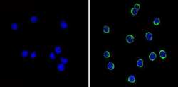
- Immunofluorescent analysis of CD44 (green) showing staining in the membrane of Hela cells (right) compared to a negative control without primary antibody (left). Formalin-fixed cells were permeabilized with 0.1% Triton X-100 in TBS for 5-10 minutes and blocked with 3% BSA-PBS for 30 minutes at room temperature. Cells were probed with a CD44 monoclonal antibody (Product # MA4405) in 3% BSA-PBS at a dilution of 1:20 and incubated overnight at 4ºC in a humidified chamber. Cells were washed with PBST and incubated with a DyLight-conjugated secondary antibody in PBS at room temperature in the dark. F-actin (red) was stained with a fluorescent red phalloidin and nuclei (blue) were stained with Hoechst or DAPI. Images were taken at a magnification of 60x.

- Submitted by
- Invitrogen Antibodies (provider)
- Main image

- Experimental details
- Immunofluorescent analysis of CD44 (green) showing staining in the membrane and cytoplasm of NIH-3T3 cells (right) compared to a negative control without primary antibody (left). Formalin-fixed cells were permeabilized with 0.1% Triton X-100 in TBS for 5-10 minutes and blocked with 3% BSA-PBS for 30 minutes at room temperature. Cells were probed with a CD44 monoclonal antibody (Product # MA4405) in 3% BSA-PBS at a dilution of 1:20 and incubated overnight at 4ºC in a humidified chamber. Cells were washed with PBST and incubated with a DyLight-conjugated secondary antibody in PBS at room temperature in the dark. F-actin (red) was stained with a fluorescent red phalloidin and nuclei (blue) were stained with Hoechst or DAPI. Images were taken at a magnification of 60x.

Supportive validation
- Submitted by
- Invitrogen Antibodies (provider)
- Main image
- Experimental details
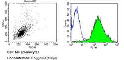
- Flow cytometry analysis of CD44 in mouse splenocytes (green) compared to an isotype control (blue). Mouse splenocytes were collected, combined with a hydrophilic polysaccharide, centrifuged, transferred to a conical tube and washed with PBS. 50 µL of cell solution was added to each tube at a dilution of 2x10^7 cells/mL, followed by the addition of 50 µL of isotype control and primary antibody (Product # MA4405) at a dilution of 0.5 µg/test. Cells were incubated for 30 min at 4ºC and washed with a cell buffer, followed by incubation with a DyLight 488-conjugated secondary antibody for 30 min at 4ºC in the dark. FACS analysis was performed using 400 µL of cell buffer.

- Submitted by
- Invitrogen Antibodies (provider)
- Main image
- Experimental details
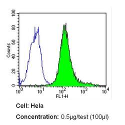
- Flow cytometry analysis of CD44 in Hela cells (green) compared to an isotype control (blue). Cells were harvested, adjusted to a concentration of 1-5x10^6 cells/mL, fixed with 2% paraformaldehyde and washed with PBS. Cells were blocked with a 2% solution of BSA-PBS for 30 min at room temperature and incubated with a CD44 monoclonal antibody (Product # MA4405) at a dilution of 0.5 µg/test for 60 min at room temperature. Cells were then incubated for 40 min at room temperature in the dark using a Dylight 488-conjugated secondary antibody and re-suspended in PBS for FACS analysis.
