 Explore
 Validate
 Learn
Learn Western blot
Antibody data
- Antibody Data
- Antigen structure
- References [2]
- Comments [0]
- Validations
- Western blot [5]
- Immunocytochemistry [1]
- Immunoprecipitation [2]
- Immunohistochemistry [2]
- Chromatin Immunoprecipitation [2]
Submit
Validation data
Reference
Comment
Report error
- Product number
- GTX110593 - Provider product page

- Provider
- GeneTex
- Proper citation
- GeneTex Cat#GTX110593, RRID:AB_11176980
- Product name
- SP1 antibody
- Antibody type
- Polyclonal
- Reactivity
- Human, Mouse, Rat
- Host
- Rabbit
Submitted references Cellular density-dependent increases in HIF-1α compete with c-Myc to down-regulate human EP4 receptor promoter activity through Sp-1-binding region.
Quercetin Reverses Rat Liver Preneoplastic Lesions Induced by Chemical Carcinogenesis.
Seira N, Yamagata K, Fukushima K, Araki Y, Kurata N, Yanagisawa N, Mashimo M, Nakamura H, Regan JW, Murayama T, Fujino H
Pharmacology research & perspectives 2018 Dec;6(6):e00441
Pharmacology research & perspectives 2018 Dec;6(6):e00441
Quercetin Reverses Rat Liver Preneoplastic Lesions Induced by Chemical Carcinogenesis.
Carrasco-Torres G, Monroy-Ramírez HC, Martínez-Guerra AA, Baltiérrez-Hoyos R, Romero-Tlalolini MLÁ, Villa-Treviño S, Sánchez-Chino X, Vásquez-Garzón VR
Oxidative medicine and cellular longevity 2017;2017:4674918
Oxidative medicine and cellular longevity 2017;2017:4674918
No comments: Submit comment
Supportive validation
- Submitted by
- GeneTex (provider)
- Main image

- Experimental details
- Sample (30 ug of whole cell lysate) A: THP-1 5% SDS PAGE GTX110593 diluted at 1:2000
- Validation comment
- WB

- Submitted by
- GeneTex (provider)
- Main image

- Experimental details
- SP1 antibody detects SP1 protein by western blot analysis. Whole cell extracts (30 ?g) was separated by 7.5% SDS-PAGE, and the membrane was blotted with SP1 antibody (GTX110593) diluted at 1:2000. The HRP-conjugated anti-rabbit IgG antibody (GTX213110-01) was used to detect the primary antibody.

- Submitted by
- GeneTex (provider)
- Main image

- Experimental details
- Non-transfected (¡V) and transfected (+) 293T whole cell extracts (50 ?g) were separated by 7.5% SDS-PAGE, and the membrane was blotted with SP1 antibody (GTX110593) diluted at 1:10000. The HRP-conjugated anti-rabbit IgG antibody (GTX213110-01) was used to detect the primary antibody, and the signal was developed with Trident ECL plus-Enhanced.

- Submitted by
- GeneTex (provider)
- Main image

- Experimental details
- Various whole cell extracts (30 ?g) were separated by 7.5% SDS-PAGE, and the membrane was blotted with SP1 antibody (GTX110593) diluted at 1:2000. The HRP-conjugated anti-rabbit IgG antibody (GTX213110-01) was used to detect the primary antibody.

Supportive validation
- Submitted by
- GeneTex (provider)
- Main image

- Experimental details
- SP1 antibody detects SP1 protein at nucleus by immunofluorescent analysis.Sample: HeLa cells were fixed in 4% paraformaldehyde at RT for 15 min.Green: SP1 protein stained by SP1 antibody (GTX110593) diluted at 1:500.Red: phalloidin, a cytoskeleton marker, diluted at 1:200.Blue: Hoechst 33342 staining.Scale bar = 10 £gm.

Supportive validation
- Submitted by
- GeneTex (provider)
- Main image

- Experimental details
- SP1 antibody immunoprecipitates SP1 protein in IP experiments. IP Sample: Raji whole cell lysate/extract A : 30 ?g whole cell lysate/extract of SP1 protein expressing Raji cells B : Control with 3 ?g of pre-immune rabbit IgG C : Immunoprecipitation of SP1 by 3 ?g of SP1 antibody (GTX110593) 5% SDS-PAGE The immunoprecipitated SP1 protein was detected by SP1 antibody (GTX110593) diluted at 1 : 1000. EasyBlot anti-rabbit IgG (HRP) (GTX221666-01) was used as a secondary reagent.
- Validation comment
- IP

- Submitted by
- GeneTex (provider)
- Main image

- Experimental details
- Immunoprecipitation of SP1 protein from THP-1 whole cell extracts using 5 £gg of OCT1 antibody (GTX110593).Western blot analysis was performed using OCT1 antibody (GTX110593).EasyBlot anti-Rabbit IgG (GTX221666-01) was used as a secondary reagent.

Supportive validation
- Submitted by
- GeneTex (provider)
- Main image
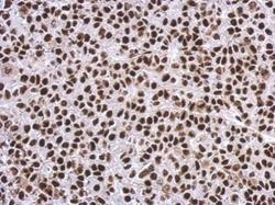
- Experimental details
- Immunohistochemical analysis of paraffin-embedded Hela xenograft, using SP1(GTX110593) antibody at 1:500 dilution.

- Submitted by
- GeneTex (provider)
- Main image

- Experimental details
- Immunohistochemical analysis of paraffin-embedded C2C12 xenograft, using SP1(GTX110593) antibody at 1:500 dilution.

Supportive validation
- Submitted by
- GeneTex (provider)
- Main image

- Experimental details
- Cross-linked ChIP was performed with 293T chromatin extract and 5 ?g of either control rabbit IgG or anti-SP1 antibody. The precipitated DNA was detected by PCR with primer set targeting to MGARP promoter.
- Validation comment
- ChIP

- Submitted by
- GeneTex (provider)
- Main image

- Experimental details
- ChIP was performed with 293T chromatin extract and 5 £gg of either normal rabbit IgG or anti-SP1 antibody. The precipitated DNA was detected by PCR with primer set targeting to MGARP promoter.
