 Explore
 Validate
 Learn
Learn Western blot
Antibody data
- Antibody Data
- Antigen structure
- References [2]
- Comments [0]
- Validations
- Western blot [4]
- Immunohistochemistry [1]
- Other assay [1]
Submit
Validation data
Reference
Comment
Report error
- Product number
- PA5-23383 - Provider product page

- Provider
- Invitrogen Antibodies
- Product name
- SOX9 Polyclonal Antibody
- Antibody type
- Polyclonal
- Antigen
- Other
- Reactivity
- Human, Mouse, Rat, Canine, Feline, Goat
- Host
- Rabbit
- Isotype
- IgG
- Vial size
- 100 µg
- Concentration
- 1 mg/mL
- Storage
- Store at 4°C short term. For long term storage, store at -20°C, avoiding freeze/thaw cycles.
Submitted references Isolation and Characterization of Multipotent Canine Urine-Derived Stem Cells.
Notch Signaling Regulates MMP-13 Expression via Runx2 in Chondrocytes.
Xu Y, Zhang T, Chen Y, Shi Q, Li M, Qin T, Hu J, Lu H, Liu J, Chen C
Stem cells international 2020;2020:8894449
Stem cells international 2020;2020:8894449
Notch Signaling Regulates MMP-13 Expression via Runx2 in Chondrocytes.
Xiao D, Bi R, Liu X, Mei J, Jiang N, Zhu S
Scientific reports 2019 Oct 30;9(1):15596
Scientific reports 2019 Oct 30;9(1):15596
No comments: Submit comment
Supportive validation
- Submitted by
- Invitrogen Antibodies (provider)
- Main image

- Experimental details
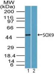
- Western blot analysis of SOX9 in human brain lysate in the 1) absence and 2) presence of immunizing peptide, 3) mouse brain and 4) rat brain lysate using a SOX9 polyclonal antibody (Product # PA5-23383) at 3 µg/mL.

- Submitted by
- Invitrogen Antibodies (provider)
- Main image
- Experimental details
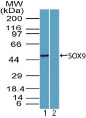
- Western blot analysis of SOX9 in mouse embryo brain lysate in the 1) absence and 2) presence of immunizing peptide. Samples were incubated in SOX9 polyclonal antibody (Product # PA5-23383) using a dilution of 2 µg/mL followed by a goat anti-rabbit Ig HRP secondary antibody. PicoTect ECL substrate solution was used for this test.
- Submitted by
- Invitrogen Antibodies (provider)
- Main image

- Experimental details
- Western blot analysis of SOX9 in human brain lysate 1) absence and 2) presence of immunizing peptide, 3) mouse brain and 4) rat brain lysate. Samples were incubated in SOX9 polyclonal antibody (Product # PA5-23383) using a dilution of 3 µg/mL.

- Submitted by
- Invitrogen Antibodies (provider)
- Main image

- Experimental details
- Western blot analysis of SOX9 in 0.5 mg/mL Human Brain lysate. Samples were incubated in SOX9 polyclonal antibody (Product # PA5-23383). This experiment was performed under reducing conditions using the 12-230 kDa separation system.

Supportive validation
- Submitted by
- Invitrogen Antibodies (provider)
- Main image
- Experimental details
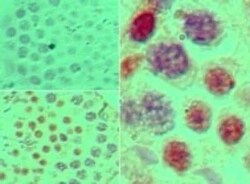
- Immunohistochemical analysis of SOX9 in formalin-fixed, paraffin-embedded mouse testis tissue. Samples were incubated in SOX9 polyclonal antibody (Product # PA5-23383) using a dilution of 5 µg/mL. Isotype control (top left) and SOX9 antibody (bottom left, right).

Supportive validation
- Submitted by
- Invitrogen Antibodies (provider)
- Main image
- Experimental details
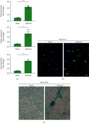
- Figure 6 Chondrogenic differentiation of urine-derived cells in vitro. (a) Chondrogenic gene (Sox9, Col2a1, Acan) expression compared between the chondrogenic medium and its respective basal cultures. * p < 0.05, ** p < 0.01. Sox9, sex-determining region Y-box 9; Col2a1, collagen type II; Acan, Aggrecan. (b) The Sox9 protein expression of the urine-derived cells after 7 days of chondrogenic induction. Scale bars = 15 mu m. (c) Alcian blue staining of cells after 21 days in chondrogenic or basal medium. Glycosaminoglycan deposition around the cells was seen in the chondrogenic induced medium. Scale bars = 20 mu m.
