 Explore
 Validate
 Learn
Learn Western blot
 Immunocytochemistry
Antibody data
- Antibody Data
- Antigen structure
- References [1]
- Comments [0]
- Validations
- Western blot [1]
Submit
Validation data
Reference
Comment
Report error
- Product number
- AF6018 - Provider product page

- Provider
- Novus Biologicals
- Product name
- Sheep Polyclonal Proprotein Convertase 2/PCSK2 Antibody
- Antibody type
- Polyclonal
- Description
- Immunogen affinity purified. Detects human Proprotein Convertase 2/PCSK2 in direct ELISAs and, human and mouse Proprotein Convertase 2/PCSK2 in Western blots.
- Reactivity
- Human, Mouse
- Host
- Sheep
- Isotype
- IgG
- Vial size
- 100 ug
- Concentration
- LYOPH
- Storage
- Use a manual defrost freezer and avoid repeated freeze-thaw cycles. 12 months from date of receipt, -20 to -70 degreesC as supplied. 1 month, 2 to 8 degreesC under sterile conditions after reconstitution. 6 months, -20 to -70 degreesC under sterile conditions after reconstitution.
Submitted references Tbr1 instructs laminar patterning of retinal ganglion cell dendrites.
Liu J, Reggiani JDS, Laboulaye MA, Pandey S, Chen B, Rubenstein JLR, Krishnaswamy A, Sanes JR
Nature neuroscience 2018 May;21(5):659-670
Nature neuroscience 2018 May;21(5):659-670
No comments: Submit comment
Supportive validation
- Submitted by
- Novus Biologicals (provider)
- Main image
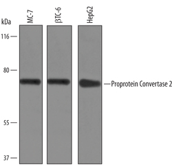
- Experimental details
- Detection of Human and Mouse Proprotein Convertase 2/PCSK2 by Western Blot. Western blot shows lysates of MCF-7 human breast cancer cell line, beta TC-6 mouse beta cell insulinoma cell line, and HepG2 human hepatocellular carcinoma cell line. PVDF Membrane was probed with 1 µg/mL of Sheep Anti-Human Proprotein Convertase 2/PCSK2 Antigen Affinity-purified Polyclonal Antibody (Catalog # AF6018) followed by HRP-conjugated Anti-Sheep IgG Secondary Antibody (Catalog # HAF016). A specific band was detected for Proprotein Convertase 2/PCSK2 at approximately 70 kDa (as indicated). This experiment was conducted under reducing conditions and using Immunoblot Buffer Group 1.
