 Explore
 Validate
 Learn
Learn Western blot
 Immunocytochemistry
 Immunohistochemistry
Antibody data
- Antibody Data
- Antigen structure
- References [4]
- Comments [0]
- Validations
- Western blot [7]
- Immunohistochemistry [3]
Submit
Validation data
Reference
Comment
Report error
- Product number
- GTX629744 - Provider product page

- Provider
- GeneTex
- Product name
- Vimentin antibody [GT812]
- Antibody type
- Monoclonal
- Reactivity
- Human, Mouse, Rat
- Host
- Mouse
Submitted references A high-throughput pipeline for validation of antibodies.
Inhibition of chronic prostate inflammation by hyaluronic acid through an immortalized human prostate stromal cell line model.
Cytokeratin 18 is necessary for initiation of TGF-β1-induced epithelial-mesenchymal transition in breast epithelial cells.
Augmented Angiogenic Factors Expression via FP Signaling Pathways in Peritoneal Endometriosis.
Sikorski K, Mehta A, Inngjerdingen M, Thakor F, Kling S, Kalina T, Nyman TA, Stensland ME, Zhou W, de Souza GA, Holden L, Stuchly J, Templin M, Lund-Johansen F
Nature methods 2018 Nov;15(11):909-912
Nature methods 2018 Nov;15(11):909-912
Inhibition of chronic prostate inflammation by hyaluronic acid through an immortalized human prostate stromal cell line model.
Liu MC, Chen WH, Chiou CS, Lo WC, Dubey NK, Chen YC, Lai WT, Yeh SD, Chiang HS, Deng WP
PloS one 2017;12(5):e0178152
PloS one 2017;12(5):e0178152
Cytokeratin 18 is necessary for initiation of TGF-β1-induced epithelial-mesenchymal transition in breast epithelial cells.
Jung H, Kim B, Moon BI, Oh ES
Molecular and cellular biochemistry 2016 Dec;423(1-2):21-28
Molecular and cellular biochemistry 2016 Dec;423(1-2):21-28
Augmented Angiogenic Factors Expression via FP Signaling Pathways in Peritoneal Endometriosis.
Rakhila H, Al-Akoum M, Doillon C, Lacroix-Pépin N, Leboeuf M, Lemyre M, Akoum A, Pouliot M
The Journal of clinical endocrinology and metabolism 2016 Dec;101(12):4752-4763
The Journal of clinical endocrinology and metabolism 2016 Dec;101(12):4752-4763
No comments: Submit comment
Enhanced validation
Supportive validation
- Submitted by
- GeneTex (provider)
- Enhanced method
- Genetic validation
- Main image
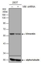
- Experimental details
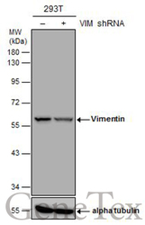
- Non-transfected (¡V) and transfected (+) 293T whole cell extracts (30 ?g) were separated by 10% SDS-PAGE, and the membrane was blotted with Vimentin antibody [GT812] (GTX629744) diluted at 1:5000.

Supportive validation
- Submitted by
- GeneTex (provider)
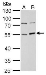
- Main image

- Experimental details
- Vimentin antibody [GT812] detects Vimentin protein by Western blot analysis.A. 30 £gg 293T whole cell lysate/extractB. 30 £gg HeLa whole cell lysate/extract10 % SDS-PAGEVimentin antibody [GT812] (GTX629744) dilution: 1:1000

- Submitted by
- GeneTex (provider)
- Main image
- Experimental details
- Vimentin antibody [GT812] detects Vimentin protein by Western blot analysis.A. 30 ?g C8D30 whole cell lysate/extractB. 30 ?g C2C12 whole cell lysate/extract10 % SDS-PAGEVimentin antibody [GT812] (GTX629744) dilution: 1:1000
- Validation comment
- WB

- Submitted by
- GeneTex (provider)
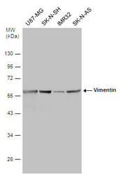
- Main image

- Experimental details
- Various whole cell extracts (30 ?g) were separated by 10% SDS-PAGE, and the membrane was blotted with Vimentin antibody [GT812] (GTX629744) diluted at 1:3000.

- Submitted by
- GeneTex (provider)
- Main image
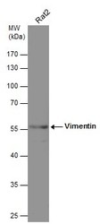
- Experimental details
- Whole cell extract (30 ?g) was separated by 10% SDS-PAGE, and the membrane was blotted with Vimentin antibody [GT812] (GTX629744) diluted at 1:75000.

- Submitted by
- GeneTex (provider)
- Main image
- Experimental details
- Various whole cell extracts (30 ?g) were separated by 10% SDS-PAGE, and the membrane was blotted with Vimentin antibody [GT812] (GTX629744) diluted at 1:5000.

- Submitted by
- GeneTex (provider)
- Main image
- Experimental details
- Non-transfected (¡V) and transfected (+) 293T whole cell extracts (30 ?g) were separated by 10% SDS-PAGE, and the membrane was blotted with Vimentin antibody [GT812] (GTX629744) diluted at 1:5000.

Supportive validation
- Submitted by
- GeneTex (provider)
- Main image
- Experimental details
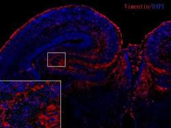
- Vimentin antibody [GT812] detects Vimentin proteins in embryonic mouse brain by immunohistochemical analysis. Sample: Frozen section of embryonic mouse brain (mE18.5). Red: Vimentin antibody [GT812] (GTX629744) diluted at 1:250. Blue: DAPI.

- Submitted by
- GeneTex (provider)
- Main image
- Experimental details
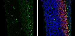
- Vimentin antibody [GT812] detects Vimentin protein expression by immunohistochemical analysis.Sample: Frozen sectioned E13.5 Rat brain. Green: Vimentin protein stained by Vimentin antibody [GT812] (GTX629744) diluted at 1:250.Red: beta Tubulin 3/ TUJ1, a mature neuron marker, stained by beta Tubulin 3/ TUJ1 antibody (GTX130245) diluted at 1:250.Blue: Fluoroshield with DAPI (GTX30920).

- Submitted by
- GeneTex (provider)
- Main image
- Experimental details
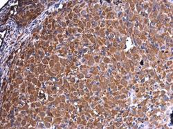
- Vimentin antibody [GT812] detects Vimentin protein at cytoplasm in rat ovary by immunohistochemical analysis. Sample: Paraffin-embedded rat ovary. Vimentin antibody [GT812] (GTX629744) diluted at 1:250.
