Explore
 Validate
 Learn
Learn Western blot
Antibody data
- Antibody Data
- Antigen structure
- References [16]
- Comments [0]
- Validations
- Western blot [3]
- Immunocytochemistry [4]
- Immunohistochemistry [2]
- Other assay [11]
Submit
Validation data
Reference
Comment
Report error
- Product number
- MA3-745 - Provider product page

- Provider
- Invitrogen Antibodies
- Product name
- Vimentin Monoclonal Antibody (J144)
- Antibody type
- Monoclonal
- Antigen
- Other
- Description
- MA3-745 detects human, rat, and mouse vimentin and has been successfully used in HELA-S3, HEK-293, rat brain, and rat kidney cells. MA3-745 has been successfully used in Western blot, immunohistochemical and immunofluorescence procedures. By Western blot, this antibody detects an ~57 kDa protein representing vimentin from HELA-S3 cells. Immunohistochemical staining of vimentin using MA3-745 can be performed. The MA3-745 immunogen was human rhabdosarcoma cell line JR1.
- Reactivity
- Human, Mouse, Rat, Canine
- Host
- Mouse
- Isotype
- IgM
- Antibody clone number
- J144
- Vial size
- 200 µL
- Concentration
- Conc. Not Determined
- Storage
- -20° C, Avoid Freeze/Thaw Cycles
Submitted references Multi-Targeting Approach in Glioblastoma Using Computer-Assisted Drug Discovery Tools to Overcome the Blood-Brain Barrier and Target EGFR/PI3Kp110β Signaling.
Leflunomide Induces Dose-Dependent Lung Injury in Mice via Stimulating Vimentin and NLRP3 Inflammasome Production.
Myocyte Enhancer Factor 2C as a New Player in Human Breast Cancer Brain Metastases.
Targeting the Temporal Dynamics of Hypoxia-Induced Tumor-Secreted Factors Halts Tumor Migration.
Establishment and characterization of a novel cell line (cc‑006cpm8) of moderately/poorly differentiated colorectal adenocarcinoma derived from a primary tumor of a patient.
Antisense oligonucleotide therapy rescues aggresome formation in a novel spinocerebellar ataxia type 3 human embryonic stem cell line.
YBX-1 mediated sorting of miR-133 into hypoxia/reoxygenation-induced EPC-derived exosomes to increase fibroblast angiogenesis and MEndoT.
Oncogenic PKC-ι activates Vimentin during epithelial-mesenchymal transition in melanoma; a study based on PKC-ι and PKC-ζ specific inhibitors.
Two novel atypical PKC inhibitors; ACPD and DNDA effectively mitigate cell proliferation and epithelial to mesenchymal transition of metastatic melanoma while inducing apoptosis.
RhoC is essential in TGF-β1 induced epithelial-mesenchymal transition in cervical cancer cells.
High-throughput subcellular protein localization using transfected-cell arrays. Subcellular protein localization using cell arrays.
Genetic deletion of COX-2 diminishes VEGF production in mouse retinal Müller cells.
Comparative proteomics profiling of a phospholamban mutant mouse model of dilated cardiomyopathy reveals progressive intracellular stress responses.
Protein composition of plasminogen activator inhibitor type 1-derived endothelial microparticles.
Isolation of lipid droplets from cells by density gradient centrifugation.
Shiga toxin binding to globotriaosyl ceramide induces intracellular signals that mediate cytoskeleton remodeling in human renal carcinoma-derived cells.
Franco C, Kausar S, Silva MFB, Guedes RC, Falcao AO, Brito MA
Cancers 2022 Jul 19;14(14)
Cancers 2022 Jul 19;14(14)
Leflunomide Induces Dose-Dependent Lung Injury in Mice via Stimulating Vimentin and NLRP3 Inflammasome Production.
El-Sherbiny M, Atef H, Eladl MA, Mohamed AS, El-Shafey M, Ali HS, Zaitone SA, Alomar SY, Alqahtani SAM, Aloyouni SY, Attia MA
Frontiers in pharmacology 2021;12:631216
Frontiers in pharmacology 2021;12:631216
Myocyte Enhancer Factor 2C as a New Player in Human Breast Cancer Brain Metastases.
Galego S, Kauppila LA, Malhó R, Pimentel J, Brito MA
Cells 2021 Feb 12;10(2)
Cells 2021 Feb 12;10(2)
Targeting the Temporal Dynamics of Hypoxia-Induced Tumor-Secreted Factors Halts Tumor Migration.
Singh M, Tian XJ, Donnenberg VS, Watson AM, Zhang J, Stabile LP, Watkins SC, Xing J, Sant S
Cancer research 2019 Jun 1;79(11):2962-2977
Cancer research 2019 Jun 1;79(11):2962-2977
Establishment and characterization of a novel cell line (cc‑006cpm8) of moderately/poorly differentiated colorectal adenocarcinoma derived from a primary tumor of a patient.
Chu X, Xue Y, Huo X, Wei J, Chen Y, Han R, Chen H, Su X, Zhang H, Gong Y, Chen J
International journal of oncology 2019 Jul;55(1):243-256
International journal of oncology 2019 Jul;55(1):243-256
Antisense oligonucleotide therapy rescues aggresome formation in a novel spinocerebellar ataxia type 3 human embryonic stem cell line.
Moore LR, Keller L, Bushart DD, Delatorre RG, Li D, McLoughlin HS, do Carmo Costa M, Shakkottai VG, Smith GD, Paulson HL
Stem cell research 2019 Aug;39:101504
Stem cell research 2019 Aug;39:101504
YBX-1 mediated sorting of miR-133 into hypoxia/reoxygenation-induced EPC-derived exosomes to increase fibroblast angiogenesis and MEndoT.
Lin F, Zeng Z, Song Y, Li L, Wu Z, Zhang X, Li Z, Ke X, Hu X
Stem cell research & therapy 2019 Aug 23;10(1):263
Stem cell research & therapy 2019 Aug 23;10(1):263
Oncogenic PKC-ι activates Vimentin during epithelial-mesenchymal transition in melanoma; a study based on PKC-ι and PKC-ζ specific inhibitors.
Ratnayake WS, Apostolatos CA, Apostolatos AH, Schutte RJ, Huynh MA, Ostrov DA, Acevedo-Duncan M
Cell adhesion & migration 2018;12(5):447-463
Cell adhesion & migration 2018;12(5):447-463
Two novel atypical PKC inhibitors; ACPD and DNDA effectively mitigate cell proliferation and epithelial to mesenchymal transition of metastatic melanoma while inducing apoptosis.
Ratnayake WS, Apostolatos AH, Ostrov DA, Acevedo-Duncan M
International journal of oncology 2017 Nov;51(5):1370-1382
International journal of oncology 2017 Nov;51(5):1370-1382
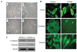
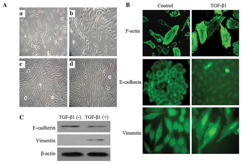
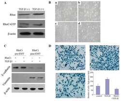
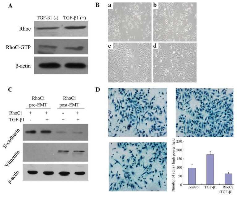
RhoC is essential in TGF-β1 induced epithelial-mesenchymal transition in cervical cancer cells.
He X, Qian Y, Cai H, Yang S, Cai J, Wang Z
Oncology letters 2015 Aug;10(2):985-989
Oncology letters 2015 Aug;10(2):985-989
High-throughput subcellular protein localization using transfected-cell arrays. Subcellular protein localization using cell arrays.
Hu Y, Janitz M
Methods in molecular biology (Clifton, N.J.) 2011;706:53-72
Methods in molecular biology (Clifton, N.J.) 2011;706:53-72
Genetic deletion of COX-2 diminishes VEGF production in mouse retinal Müller cells.
Yanni SE, McCollum GW, Penn JS
Experimental eye research 2010 Jul;91(1):34-41
Experimental eye research 2010 Jul;91(1):34-41
Comparative proteomics profiling of a phospholamban mutant mouse model of dilated cardiomyopathy reveals progressive intracellular stress responses.
Gramolini AO, Kislinger T, Alikhani-Koopaei R, Fong V, Thompson NJ, Isserlin R, Sharma P, Oudit GY, Trivieri MG, Fagan A, Kannan A, Higgins DG, Huedig H, Hess G, Arab S, Seidman JG, Seidman CE, Frey B, Perry M, Backx PH, Liu PP, MacLennan DH, Emili A
Molecular & cellular proteomics : MCP 2008 Mar;7(3):519-33
Molecular & cellular proteomics : MCP 2008 Mar;7(3):519-33
Protein composition of plasminogen activator inhibitor type 1-derived endothelial microparticles.
Sander TL, Ou JS, Densmore JC, Kaul S, Matus I, Twigger S, Halligan B, Greene AS, Pritchard KA Jr, Oldham KT
Shock (Augusta, Ga.) 2008 Apr;29(4):504-11
Shock (Augusta, Ga.) 2008 Apr;29(4):504-11
Isolation of lipid droplets from cells by density gradient centrifugation.
Brasaemle DL, Wolins NE
Current protocols in cell biology 2006 Jan;Chapter 3:Unit 3.15
Current protocols in cell biology 2006 Jan;Chapter 3:Unit 3.15
Shiga toxin binding to globotriaosyl ceramide induces intracellular signals that mediate cytoskeleton remodeling in human renal carcinoma-derived cells.
Takenouchi H, Kiyokawa N, Taguchi T, Matsui J, Katagiri YU, Okita H, Okuda K, Fujimoto J
Journal of cell science 2004 Aug 1;117(Pt 17):3911-22
Journal of cell science 2004 Aug 1;117(Pt 17):3911-22
No comments: Submit comment
Supportive validation
- Submitted by
- Invitrogen Antibodies (provider)
- Main image

- Experimental details
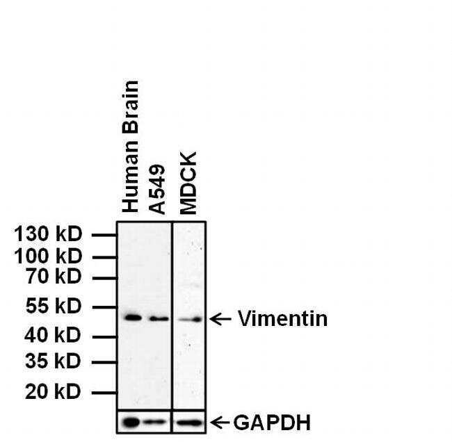
- Western blot analysis of Vimentin was performed by loading 20 µg of indicated whole cell lysates and 7 µL of PageRuler Prestained Protein Ladder (Product # 26616) per well onto a 4-20% Tris-Glycine polyacrylamide gel (Product # WT4202BX10). Proteins were transferred to a nitrocellulose membrane using the G2 Blotter (Product # 62288), and blocked with 5% Milk in TBST for 1 hour at room temperature. Vimentin was detected at ~53 kDa using a Vimentin mouse monoclonal antibody (Product # MA3-745) at a dilution of 1:500 in blocking buffer overnight at 4°C on a rocking platform. GAPDH was detected using GAPDH rabbit polyclonal antibody, HRP conjugate (Product # PA1-987-HRP) overnight 4°C on a rocking platform. Chemiluminescent detection was performed using SuperSignal West Pico (Product # 34078).
- Submitted by
- Invitrogen Antibodies (provider)
- Main image

- Experimental details
- Western blot analysis of Vimentin was performed by loading 20 µg of indicated whole cell lysates and 7 µL of PageRuler Prestained Protein Ladder (Product # 26616) per well onto a 4-20% Tris-Glycine polyacrylamide gel (Product # WT4202BX10). Proteins were transferred to a nitrocellulose membrane using the G2 Blotter (Product # 62288), and blocked with 5% Milk in TBST for 1 hour at room temperature. Vimentin was detected at ~53 kDa using a Vimentin mouse monoclonal antibody (Product # MA3-745) at a dilution of 1:500 in blocking buffer overnight at 4°C on a rocking platform. GAPDH was detected using GAPDH rabbit polyclonal antibody, HRP conjugate (Product # PA1-987-HRP) overnight 4°C on a rocking platform. Chemiluminescent detection was performed using SuperSignal West Pico (Product # 34078).

- Submitted by
- Invitrogen Antibodies (provider)
- Main image
- Experimental details
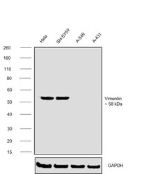
- Western blot was performed using Anti-Vimentin Monoclonal Antibody (J144) (Product # MA3-745) and a ~58 kDa band corresponding to VIM was observed across cell lines tested . Whole cell extracts (30 µg lysate) of HeLa (Lane 1), SH-SY5Y (Lane 2), A549 (Lane 3), A-431 (Lane 4) were electrophoresed using NuPAGE™ 4-12% Bis-Tris Protein Gel (Product # NP0321BOX). Resolved proteins were then transferred onto a nitrocellulose membrane (Product # IB23001) by iBlot® 2 Dry Blotting System (Product # IB21001). The blot was probed with the primary antibody (1:1000) and detected by chemiluminescence with Goat anti-Mouse IgG (H+L) Superclonal™ Recombinant Secondary Antibody, HRP (Product # A28177,1:20000) using the iBright™ FL1500 Imaging System (Product # A44115). Chemiluminescent detection was performed using SuperSignal™ West Pico PLUS Chemiluminescent Substrate (Product # 34580).

Supportive validation
- Submitted by
- Invitrogen Antibodies (provider)
- Main image
- Experimental details
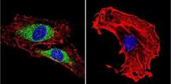
- Immunofluorescent analysis of Vimentin using Vimentin Monoclonal antibody (J144) (Product # MA3-745) shows staining in HEK293 cells. Vimentin staining (green), F-Actin staining with Phalloidin (red) and nuclei with DAPI (blue) is shown. Cells were grown on chamber slides and fixed with formaldehyde prior to staining. Cells were probed without (control) or with or an antibody recognizing Vimentin (Product # MA3-745) at a dilution of 1:100-1:200 over night at 4 °C, washed with PBS and incubated with a DyLight-488 conjugated secondary antibody (Product # 35552 for GAR, Product # 35503 for GAM). Images were taken at 60X magnification.
- Submitted by
- Invitrogen Antibodies (provider)
- Main image
- Experimental details
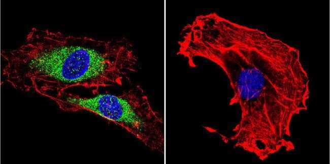
- Immunofluorescent analysis of Vimentin using Vimentin Monoclonal antibody (J144) (Product # MA3-745) shows staining in C6 glioma cells. Vimentin staining (green), F-Actin staining with Phalloidin (red) and nuclei with DAPI (blue) is shown. Cells were grown on chamber slides and fixed with formaldehyde prior to staining. Cells were probed without (control) or with or an antibody recognizing Vimentin (Product # MA3-745) at a dilution of 1:100-1:200 over night at 4 °C, washed with PBS and incubated with a DyLight-488 conjugated secondary antibody (Product # 35552 for GAR, Product # 35503 for GAM). Images were taken at 60X magnification.
- Submitted by
- Invitrogen Antibodies (provider)
- Main image
- Experimental details
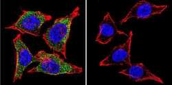
- Immunofluorescent analysis of Vimentin using Vimentin Monoclonal antibody (J144) (Product # MA3-745) shows staining in HeLa cells. Vimentin staining (green), F-Actin staining with Phalloidin (red) and nuclei with DAPI (blue) is shown. Cells were grown on chamber slides and fixed with formaldehyde prior to staining. Cells were probed without (control) or with or an antibody recognizing Vimentin (Product # MA3-745) at a dilution of 1:100-1:200 over night at 4 °C, washed with PBS and incubated with a DyLight-488 conjugated secondary antibody (Product # 35552 for GAR, Product # 35503 for GAM). Images were taken at 60X magnification.

- Submitted by
- Invitrogen Antibodies (provider)
- Main image
- Experimental details
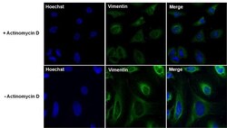
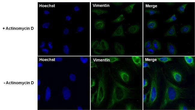
- Immunofluorescent analysis of Vimentin (green) in HeLa cells untreated or treated with 1uM Actinomycin D for 19 hours. The cells were fixed with 4% Paraformaldehyde for 15 minutes, permeabilized with 0.1% Triton X-100 for 10 minutes, and blocked with 3% BSA for 30 minutes at room temperature. Cells were stained with a Vimentin mouse monoclonal antibody (Product # MA3-745) at a dilution of 1:100 in blocking buffer for 1 hour at room temperature, and then incubated with a Goat anti-Mouse IgG (H+L) Secondary Antibody, Alexa Fluor Plus 488 conjugate (Product # A32723) at a dilution of 1:500 for at least 30 minutes at a room temperature in the dark (green). Nuclei (blue) were stained with Hoechst 33342 (Product # 62249). Images were taken on a Thermo Scientific ToxInsight Instrument at 20X magnification.
Supportive validation
- Submitted by
- Invitrogen Antibodies (provider)
- Main image

- Experimental details
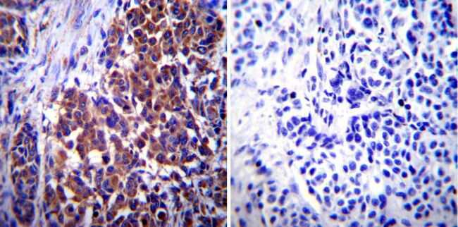
- Immunohistochemistry was performed on biopsies of deparaffinized Human melanoma tissue. To expose target proteins, heat induced antigen retrieval was performed using 10mM sodium citrate (pH6.0) buffer, microwaved for 8-15 minutes. Following antigen retrieval tissues were blocked in 3% BSA-PBS for 30 minutes at room temperature. Tissues were then probed at a dilution of 1:1000 with a mouse monoclonal antibody recognizing Vimentin (Product # MA3-745) or without primary antibody (negative control) overnight at 4°C in a humidified chamber. Tissues were washed extensively with PBST and endogenous peroxidase activity was quenched with a peroxidase suppressor. Detection was performed using a biotin-conjugated secondary antibody and SA-HRP, followed by colorimetric detection using DAB. Tissues were counterstained with hematoxylin and prepped for mounting.
- Submitted by
- Invitrogen Antibodies (provider)
- Main image

- Experimental details
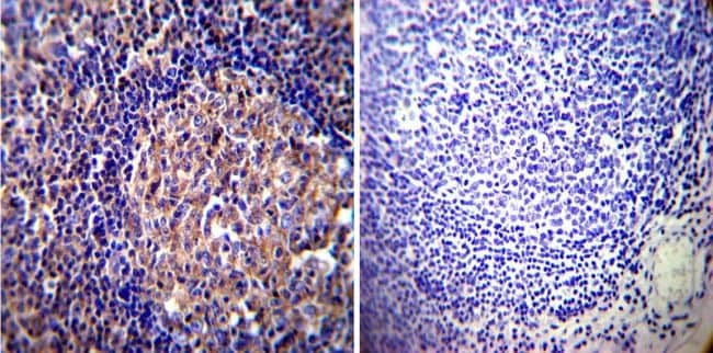
- Immunohistochemistry was performed on both normal and cancer biopsies of deparaffinized Human tonsil tissue. To expose target proteins, heat induced antigen retrieval was performed using 10mM sodium citrate (pH6.0) buffer, microwaved for 8-15 minutes. Following antigen retrieval tissues were blocked in 3% BSA-PBS for 30 minutes at room temperature. Tissues were then probed at a dilution of 1:1000 with a mouse monoclonal antibody recognizing Vimentin (Product # MA3-745) or without primary antibody (negative control) overnight at 4°C in a humidified chamber. Tissues were washed extensively with PBST and endogenous peroxidase activity was quenched with a peroxidase suppressor. Detection was performed using a biotin-conjugated secondary antibody and SA-HRP, followed by colorimetric detection using DAB. Tissues were counterstained with hematoxylin and prepped for mounting.
Supportive validation
- Submitted by
- Invitrogen Antibodies (provider)
- Main image
- Experimental details
- NULL
- Submitted by
- Invitrogen Antibodies (provider)
- Main image
- Experimental details
- NULL
- Submitted by
- Invitrogen Antibodies (provider)
- Main image

- Experimental details
- Figure 8 PKC-iota strongly associates with vimentin. Whole cell lysates (100 u g) of malignant cells (Sk-MeL-2 and MeWo) were IP separately for PKC-iota and vimentin using specific antibodies. First column of the western blot analysis represents the (+) control which contained 40 u g of MeWo whole cell extract, applied to ensure that bands appeared for the specific proteins in western blots. Western blots of PKC-iota IP showed an association with vimentin while no association was observed for E-cadherin, CD44 and NF-kappaB p65. Vimentin IP confirmed the association with PKC-iota the western blot while no association was observed with above mentioned proteins. Three experiments were performed in each trial. Densitometry for each band is indicated in the bar graph.

- Submitted by
- Invitrogen Antibodies (provider)
- Main image

- Experimental details
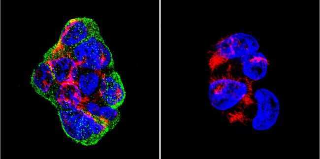
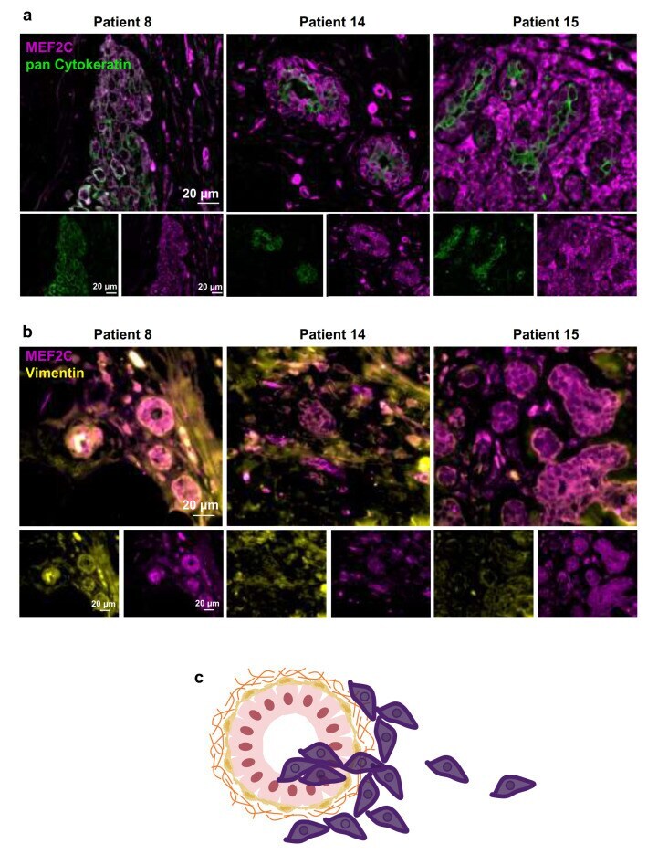
- Figure 2 Myocyte Enhancer Factor 2C expression in human breast cancer primary tumors. Double immunofluorescence analysis of myocyte enhancer factor 2C (MEF2C; purple) with the epithelial and tumoral marker, pan Cytokeratin (green), showed that MEF2C expressing cells did not significantly express pan Cytokeratin and were found in disorganized mammary ducts as well as in the surrounding tissue ( a ). Double immunofluorescence analysis of MEF2C (purple) with the mesenchymal marker, vimentin (yellow), showed that MEF2C expressing cells in mammary ducts also expressed vimentin ( b ). Schematic representation of the first stages of the metastatic cascade, showing MEF2C expressing cells (purple) in the mammary duct and invading the surrounding tissue ( c ). Three resected human primary BC cases were studied; ten fields per section and one section per case were analyzed.
- Submitted by
- Invitrogen Antibodies (provider)
- Main image
- Experimental details
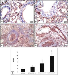
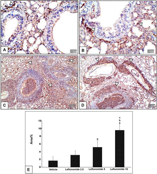
- FIGURE 8 Immunohistochemical staining of vimentin in lung sections (A) An image from the vehicle group shows weak expression of vimentin compared to moderately increased expression of vimentin in mesenchymal cells in inter alveolar septa, perivascular and peribronchiolar spaces in leflunomide 2.5 group (B) , leflunomide 5 group (C) and markedly increased in leflunomide 10 group (D) (E) A column chart representing the mean area +- SDM, (A) : vs. vehicle group, (B) : vs. the leflunomide 2.5 mg per kg group, (C) : vs. the leflunomide 5 mg per kg group at p < 0.05.
- Submitted by
- Invitrogen Antibodies (provider)
- Main image

- Experimental details
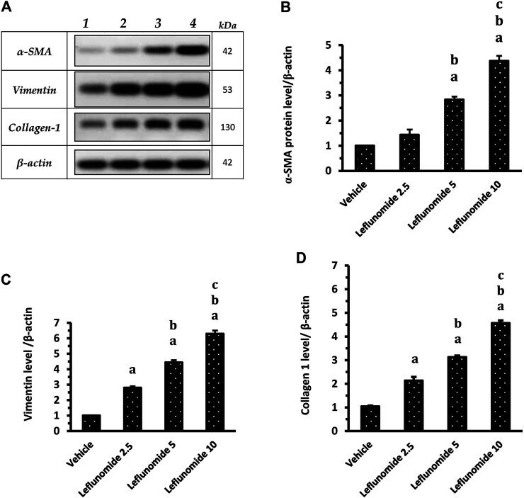
- FIGURE 10 Western blot analysis for alpha-SMA, vimentin and collagen 1 proteins. (A) : Western blot gels of the target genes in (1) vehicle group, (2) leflunomide 2.5 mg/kg group, (3) leflunomide 5 mg/kg group and (4) leflunomide 10 mg/kg group. ( B-D) : Column charts , vimentin and collagen 1 proteins relative to beta-actin. The quantification data are presented as mean +- SDM. (A) : vs. the vehicle group, (B) : vs. leflunomide 2.5 mg per kg group, (C) : vs. leflunomide 5 mg per kg group at p < 0.05. alpha-SMA: alpha-smooth muscle actin.
- Submitted by
- Invitrogen Antibodies (provider)
- Main image

- Experimental details
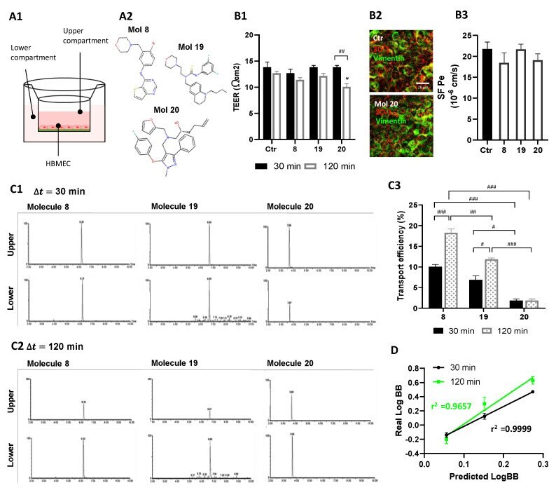
- Selected molecules proved to efficiently cross the BBB. ( A ) HBMEC were grown to confluence in Transwell inserts and then incubated with molecules 8, 19, or 20 at U87MG EC 50 , or vehicle (control; Ctr), for 30 or 120 min. After that, ( B ) inserts were removed and tested for barrier integrity, and ( C ) cell medium from upper and lower chambers were collected and analyzed by UPLC-MS/MS to assess molecule permeation across HBMEC. ( A1 ) Schematic view of the two-chamber BBB model and ( A2 ) molecular structure of the candidates tested for BBB permeation. ( B1 ) BBB integrity was assessed by TEER measurement. ( B2 ) Representative immunostaining for HBMEC expression of ZO-1 and vimentin, junctional, and cytoskeleton proteins, respectively. ( B3 ) Quantitative analysis of the permeability to SF (SF Pe) across HBMEC in the Transwell system. ( C ) Total Ion Chromatograms (TIC) of MRM analyses obtained by UPLC-MS/MS. Quantification was based on the integration (peak areas) of the represented well-resolved peaks with reproducible retention times, both in upper and lower compartments at ( C1 ) 30 min and ( C2 ) 120 min of incubation with molecule 8, 19, or 20. ( C3 ) Transport efficiency quantification is expressed as a percentage of initial applied concentration. ( D ) Linear correlation between predicted and experimentally validated values of LogBB, expressing the degree of BBB permeation. Statistical analysis was performed by one-way ANOVA with Tukey correction. Data presented i
- Submitted by
- Invitrogen Antibodies (provider)
- Main image
- Experimental details
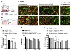
- Figure 11 A reliable co-culture system corroborated candidate molecules as BBB permeable and strong anti-GB agents: ( A ) HBMEC were grown to confluence in Transwell inserts and U87MG or U87MG-wtEGFR cells were seeded in the lower chamber. After 3 days of co-culture, supernatant in the upper chamber was replaced with fresh medium (control) or with medium supplemented with molecules 8, 19, or 20 at EC 50 . After 2 h, the inserts were removed and tested for barrier integrity ( B - D ), whereas the gliomasphere-like cells were maintained in culture for more than 24 h to assess cell viability by MTT assay ( E ). In parallel, the U87MG or U87MG-wtEGFR cells were incubated for 24 h with the basolateral conditioned medium of HBMEC after incubation with the molecules. ( A1 ) Schematic representation of the BBB-GB co-culture model and ( A2 ) of the conditioned medium assay. ( B ) Immunostaining for ZO-1 and vimentin in HBMEC mono-culture or co-cultured with U87MG or U87MG-wtEGFR. Images are representative of three independent experiments each with 10 random fields analyzed. ( C ) Semi-quantitative analysis of ZO-1 expression in HBMEC cells mono-cultured vs. co-cultured with U87MG or U87MG-wtEGFR by mean fluorescence intensity fold-change. ( D ) TEER of HBMEC after 2 h of incubation with the molecules both in mono- or co-culture system. ( E ) U87MG or U87MG-wtEGFR cell viability assessment by MTT assay after molecules exposure either in co-culture system or by conditioned medium. Data
- Submitted by
- Invitrogen Antibodies (provider)
- Main image

- Experimental details
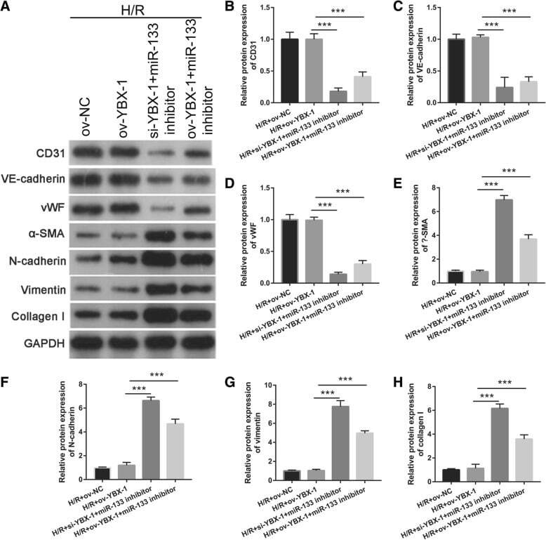
- Fig. 10 Intercellular transfer of miR-133 by H/R-induced EPC-derived exosomes inhibits fibroblast MEndoT. a Endothelial markers CD31, VE-cadherin, and vWF and fibrosis markers alpha-SMA, N-cadherin, vimentin, and collagen I were measured by western blotting in fibroblasts treated with H/R, H/R+ov-NC, H/R+si-YBX1+miR-133 inhibitor, and H/R+ ov-YBX1+miR-133 inhibitor-induced EPC-derived exosomes. b - h The bar graph represents quantification of endothelial markers CD31 ( b ), VE-cadherin ( c ), and vWF ( d ) and fibrosis markers alpha-SMA ( e ), N-cadherin ( f ), vimentin ( g ), and collagen I ( h ) expression per group. *** P < 0.001
- Submitted by
- Invitrogen Antibodies (provider)
- Main image

- Experimental details
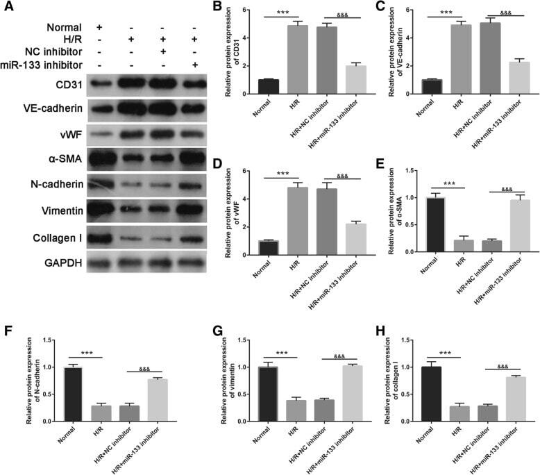
- Fig. 5 Intercellular transfer of miR-133 by H/R-induced EPC-derived exosomes inhibits fibroblast MEndoT. a Endothelial markers CD31, VE-cadherin, and vWF and fibrosis markers alpha-SMA, N-cadherin, vimentin, and collagen I were measured by western blotting in fibroblasts treated with normal cultured EPC-derived exosomes, H/R-induced EPC-derived exosomes, H/R+NC inhibitor-induced EPC-derived exosomes, and H/R+miR-133 inhibitor-induced EPC-derived exosomes. b - h The bar graph represents quantification of endothelial markers CD31 ( b ), VE-cadherin ( c ), and Vwf ( d ) and fibrosis markers alpha-SMA ( e ), N-cadherin ( f ), vimentin ( g ), and collagen I ( h ) expression per group. *** P < 0.001, &&& P < 0.001
- Submitted by
- Invitrogen Antibodies (provider)
- Main image

- Experimental details
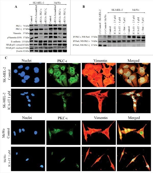
- Figure 6. PKC-iota activates Vimentin and both aPKCs stimulate nuclei translocation of NF-kappaB in melanoma cells. The effects of on si RNA knockdown (20 nM of PKC-iota and PKC-zeta si RNA) on the expression of aPKCs, Vimentin activation and NF-kappaB translocation are shown in Fig. 6A . 40 ug of protein was loaded in to each well and beta-actin was used as the loading control in each Western blot. Fig. 6B is shown the association between PKC-iota and Par6. Whole cell lysates (100 ug) of malignant cells (Sk-Mel-2 and MeWo) were immunoprecipitated separately for PKC-iota and Par6 using specific antibodies. First two lanes in Western blot represents the (+) control which contained 40 ug of Sk-MEL-2 and MeWo whole cell extracts, respectively, applied to ensure that bands appeared for the specific proteins in Western blots. Western blots of PKC-iota immunoprecipitation showed an association with Par6. Reverse-immunoprecipitation of Par6 confirmed the association with PKC-iota while no association was observed with PKC-zeta. Fig. 6C represents the immunofluorescence staining of nuclei (blue panel), PKC-iota (green panel) and Vimentin (red panel) for melanoma cells (SK-MEL-2 and MeWo) treated with ICA-1T (1 muM) against controls. The images were captured at 200X magnification. Experiments ( N = 3) were performed in each trial.