 Explore
 Validate
 Learn
Learn Western blot
 Immunocytochemistry
Antibody data
- Antibody Data
- Antigen structure
- References [4]
- Comments [0]
- Validations
- Western blot [7]
- Immunoprecipitation [1]
- Immunohistochemistry [6]
Submit
Validation data
Reference
Comment
Report error
- Product number
- NBP1-31327 - Provider product page

- Provider
- Novus Biologicals
- Proper citation
- Novus Cat#NBP1-31327, RRID:AB_10003780
- Product name
- Rabbit Polyclonal Vimentin Antibody
- Antibody type
- Polyclonal
- Description
- Immunogen affinity purified.
- Reactivity
- Human, Mouse, Rat
- Host
- Rabbit
- Isotype
- IgG
- Vial size
- 0.1 ml
- Storage
- Aliquot and store at -20C or -80C. Avoid freeze-thaw cycles.
Submitted references VCAM-1 secreted from cancer-associated fibroblasts enhances the growth and invasion of lung cancer cells through AKT and MAPK signaling.
Extracellular vesicles secreted by hypoxia pre-challenged mesenchymal stem cells promote non-small cell lung cancer cell growth and mobility as well as macrophage M2 polarization via miR-21-5p delivery.
Probing Functional Changes in Exocyst Configuration with Monoclonal Antibodies.
Amniotic fluid proteome analysis from Down syndrome pregnancies for biomarker discovery.
Zhou Z, Zhou Q, Wu X, Xu S, Hu X, Tao X, Li B, Peng J, Li D, Shen L, Cao Y, Yang L
Cancer letters 2020 Mar 31;473:62-73
Cancer letters 2020 Mar 31;473:62-73
Extracellular vesicles secreted by hypoxia pre-challenged mesenchymal stem cells promote non-small cell lung cancer cell growth and mobility as well as macrophage M2 polarization via miR-21-5p delivery.
Ren W, Hou J, Yang C, Wang H, Wu S, Wu Y, Zhao X, Lu C
Journal of experimental & clinical cancer research : CR 2019 Feb 8;38(1):62
Journal of experimental & clinical cancer research : CR 2019 Feb 8;38(1):62
Probing Functional Changes in Exocyst Configuration with Monoclonal Antibodies.
Inamdar SM, Hsu SC, Yeaman C
Frontiers in cell and developmental biology 2016;4:51
Frontiers in cell and developmental biology 2016;4:51
Amniotic fluid proteome analysis from Down syndrome pregnancies for biomarker discovery.
Cho CK, Smith CR, Diamandis EP
Journal of proteome research 2010 Jul 2;9(7):3574-82
Journal of proteome research 2010 Jul 2;9(7):3574-82
No comments: Submit comment
Supportive validation
- Submitted by
- Novus Biologicals (provider)
- Main image

- Experimental details
- Western Blot: Vimentin Antibody [NBP1-31327] - 293T cell lysate: A. 20 ug. B. 10 ug. C. 5 ug. D. 1 ug.

- Submitted by
- Novus Biologicals (provider)
- Main image

- Experimental details
- Western Blot: Vimentin Antibody [NBP1-31327] - Various whole cell extracts (30 ug) were separated by 10% SDS-PAGE, and the membrane was blotted with Vimentin antibody diluted at 1:10000. The HRP-conjugated anti-rabbit IgG antibody was used to detect the primary antibody.
- Submitted by
- Novus Biologicals (provider)
- Main image
- Experimental details
- Western Blot: Vimentin Antibody [NBP1-31327] - Whole cell extracts (30 ug) was separated by 10% SDS-PAGE, and the membrane was blotted with Vimentin antibody at a dilution of 1:10000. The HRP-conjugated anti-rabbit IgG antibody was used to detect the primary antibody.

- Submitted by
- Novus Biologicals (provider)
- Main image
- Experimental details
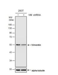
- Western Blot: Vimentin Antibody [NBP1-31327] - Non-transfected (-) and transfected (+) 293T whole cell extracts (30 ug) were separated by 10% SDS-PAGE, and the membrane was blotted with Vimentin antibody.

- Submitted by
- Novus Biologicals (provider)
- Main image

- Experimental details
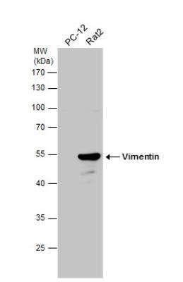
- Western Blot: Vimentin Antibody [NBP1-31327] - Various whole cell extracts (30 ug) were separated by 10% SDS-PAGE, membrane was blotted with Vimentin antibody at 1:50000.

- Submitted by
- Novus Biologicals (provider)
- Main image

- Experimental details
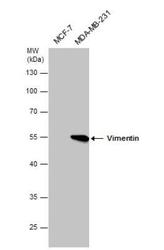
- Western Blot: Vimentin Antibody [NBP1-31327] - Various whole cell extracts (30 ug) were separated by 10% SDS-PAGE, membrane was blotted with Vimentin antibody at 1:50000.

- Submitted by
- Novus Biologicals (provider)
- Main image

- Experimental details
- Western Blot: Vimentin Antibody [NBP1-31327] - Whole cell extract (30 ug) was separated by 10% SDS-PAGE, and the membrane was blotted with Vimentin antibody diluted at 1:2000.

Supportive validation
- Submitted by
- Novus Biologicals (provider)
- Main image

- Experimental details
- Immunoprecipitation: Vimentin Antibody [NBP1-31327] - Vimentin protein from HeLa whole cell extracts using 5 ug of Vimentin antibody. Western blot analysis was performed using Vimentin antibody. EasyBlot anti-Rabbit IgG was used as a secondary reagent.

Supportive validation
- Submitted by
- Novus Biologicals (provider)
- Main image
- Experimental details
- Immunohistochemistry-Paraffin: Vimentin Antibody [NBP1-31327] - Paraffin-embedded U37C xenograft, using antibody at 1:500 dilution.

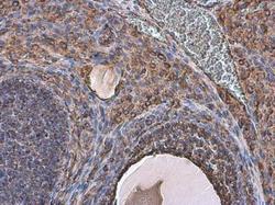
- Submitted by
- Novus Biologicals (provider)
- Main image
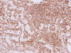
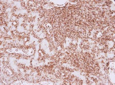
- Experimental details
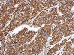
- Immunohistochemistry-Paraffin: Vimentin Antibody [NBP1-31327] - Paraffin-embedded human lung adenocarcinoma. Vimentin antibody diluted at 1:500.
- Submitted by
- Novus Biologicals (provider)
- Main image
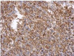
- Experimental details
- Immunohistochemistry-Paraffin: Vimentin Antibody [NBP1-31327] - Analysis of paraffin-embedded RT2 xenograft, using Vimentin antibody at 1:500 dilution.

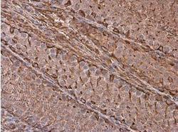
- Submitted by
- Novus Biologicals (provider)
- Main image
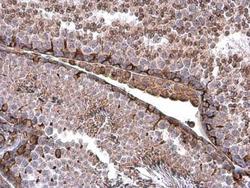
- Experimental details
- Immunohistochemistry-Paraffin: Vimentin Antibody [NBP1-31327] - Paraffin-embedded mouse testis. Vimentin antibody at 1:500.
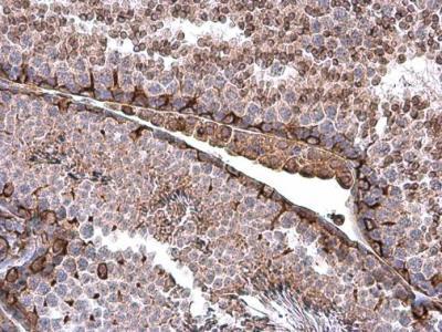
- Submitted by
- Novus Biologicals (provider)
- Main image
- Experimental details
- Immunohistochemistry-Paraffin: Vimentin Antibody [NBP1-31327] - Paraffin-embedded rat testis. Vimentin antibody at 1:500.

- Submitted by
- Novus Biologicals (provider)
- Main image
- Experimental details
- Immunohistochemistry-Paraffin: Vimentin Antibody [NBP1-31327] - Paraffin-embedded rat ovary. Vimentin antibody at 1:500.
