Explore
 Validate
 Learn
Learn Western blot
 Immunohistochemistry
Antibody data
- Antibody Data
- Antigen structure
- References [7]
- Comments [0]
- Validations
- Western blot [3]
- Immunocytochemistry [2]
- Other assay [4]
Submit
Validation data
Reference
Comment
Report error
- Product number
- PA1-16759 - Provider product page

- Provider
- Invitrogen Antibodies
- Product name
- Vimentin Polyclonal Antibody
- Antibody type
- Polyclonal
- Antigen
- Purifed from natural sources
- Reactivity
- Human, Mouse, Rat, Bovine, Canine, Chicken/Avian, Porcine
- Host
- Chicken/Avian
- Isotype
- IgY
- Vial size
- 100 µL
- Concentration
- 16.6 mg/mL
- Storage
- Store at 4°C short term. For long term storage, store at -20°C, avoiding freeze/thaw cycles.
Submitted references RhoA drives actin compaction to restrict axon regeneration and astrocyte reactivity after CNS injury.
Biomimetic electromechanical stimulation to maintain adult myocardial slices in vitro.
ROCK2 inhibition triggers the collective invasion of colorectal adenocarcinomas.
4-methylumbelliferone Prevents Liver Fibrosis by Affecting Hyaluronan Deposition, FSTL1 Expression and Cell Localization.
The LepR-mediated leptin transport across brain barriers controls food reward.
Tanycytes control the hormonal output of the hypothalamic-pituitary-thyroid axis.
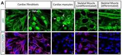
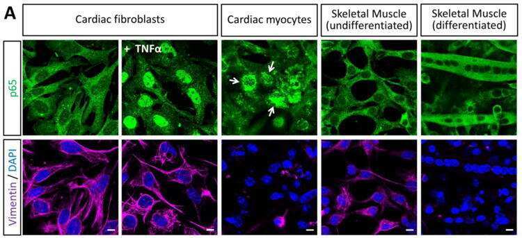
NF-κB activation is cell type-specific in the heart.
Stern S, Hilton BJ, Burnside ER, Dupraz S, Handley EE, Gonyer JM, Brakebusch C, Bradke F
Neuron 2021 Nov 3;109(21):3436-3455.e9
Neuron 2021 Nov 3;109(21):3436-3455.e9
Biomimetic electromechanical stimulation to maintain adult myocardial slices in vitro.
Watson SA, Duff J, Bardi I, Zabielska M, Atanur SS, Jabbour RJ, Simon A, Tomas A, Smolenski RT, Harding SE, Perbellini F, Terracciano CM
Nature communications 2019 May 15;10(1):2168
Nature communications 2019 May 15;10(1):2168
ROCK2 inhibition triggers the collective invasion of colorectal adenocarcinomas.
Libanje F, Raingeaud J, Luan R, Thomas Z, Zajac O, Veiga J, Marisa L, Adam J, Boige V, Malka D, Goéré D, Hall A, Soazec JY, Prall F, Gelli M, Dartigues P, Jaulin F
The EMBO journal 2019 Jul 15;38(14):e99299
The EMBO journal 2019 Jul 15;38(14):e99299
4-methylumbelliferone Prevents Liver Fibrosis by Affecting Hyaluronan Deposition, FSTL1 Expression and Cell Localization.
Andreichenko IN, Tsitrina AA, Fokin AV, Gabdulkhakova AI, Maltsev DI, Perelman GS, Bulgakova EV, Kulikov AM, Mikaelyan AS, Kotelevtsev YV
International journal of molecular sciences 2019 Dec 13;20(24)
International journal of molecular sciences 2019 Dec 13;20(24)
The LepR-mediated leptin transport across brain barriers controls food reward.
Di Spiezio A, Sandin ES, Dore R, Müller-Fielitz H, Storck SE, Bernau M, Mier W, Oster H, Jöhren O, Pietrzik CU, Lehnert H, Schwaninger M
Molecular metabolism 2018 Feb;8:13-22
Molecular metabolism 2018 Feb;8:13-22
Tanycytes control the hormonal output of the hypothalamic-pituitary-thyroid axis.
Müller-Fielitz H, Stahr M, Bernau M, Richter M, Abele S, Krajka V, Benzin A, Wenzel J, Kalies K, Mittag J, Heuer H, Offermanns S, Schwaninger M
Nature communications 2017 Sep 7;8(1):484
Nature communications 2017 Sep 7;8(1):484
NF-κB activation is cell type-specific in the heart.
Rivera-Serrano EE, Sherry B
Virology 2017 Feb;502:133-143
Virology 2017 Feb;502:133-143
No comments: Submit comment
Supportive validation
- Submitted by
- Invitrogen Antibodies (provider)
- Main image
- Experimental details
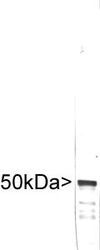
- Western blot of crude extract of human embryonic kidney Hek293 cells stained with PA1-16759, showing a single strong clean band at ~50kDa.
- Submitted by
- Invitrogen Antibodies (provider)
- Main image
- Experimental details
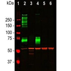
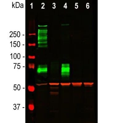
- Western blot analysis of Vimentin in tissue and cell lysates. Samples were incubated in Vimentin polyclonal antibody (Product # PA1-16759 using a dilution of 1:5000. Antibody in red. [1] protein standard (red), [2] rat whole brain lysate, [3] HeLa, [4] SH-SY5Y, [5] HEK293, and [6] NIH-3T3 cell lysates. NB300-223 binds to the vimentin protein showing a single band at ~50 kDa. The blot was simultaneously probed with mouse mAb to MAP2C/D, dilution 1:5000 in green, revealing multiple bands around 280 kDa that correspond to full length MAP2A/2B isotypes, and ~70 kDa bands which are MAP2C/D isotypes. MAP2 isotypes are seen only in extracts containing neuronal lineage cells.
- Submitted by
- Invitrogen Antibodies (provider)
- Main image
- Experimental details
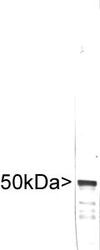
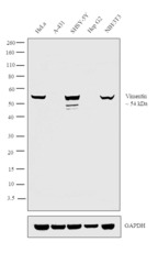
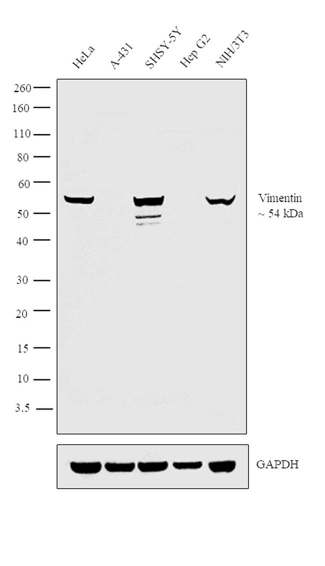
- Western blot analysis was performed on Whole cell extracts (30 µg lysate) of HeLa (Lane 1), A-431 (Lane 2), SHSY-5Y (Lane 3), Hep G2 (Lane 4) and NIH/3T3 (Lane 5). The blot was probed with Anti-Vimentin Polyclonal Antibody (Product # PA1-16759, 1:2000 dilution) and detected by chemiluminescence using Goat anti-Chicken IgY (H+L) Secondary Antibody, HRP (Product # A16054, 0.25 µg/ml, 1:4000 dilution). A 54 kDa band corresponding to Vimentin was observed across all the cell lines positive for Vimentin (Lanes 1, 3 and 5), while this band was absent in the cell lines which do not express Vimentin protein (Lanes 2 and 4).
Supportive validation
- Submitted by
- Invitrogen Antibodies (provider)
- Main image

- Experimental details
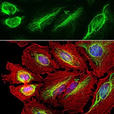
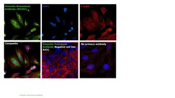
- Immunocytochemistry analysis of Vimentin in HeLa cells. Samples were incubated in Vimentin polyclonal antibody (Product # PA1-16759) using a dilution of 1:10000. Antibody in green and costained with mouse mAb to actin, dilution 1:500 in red. The blue is DAPI staining of nuclear DNA. The vimentin antibody stains the intermediate filament network while the actin antibody labels the submembranous cytoskeleton, stress fibers, and bundles of actin associated with cell adhesion sites.
- Submitted by
- Invitrogen Antibodies (provider)
- Main image
- Experimental details
- Immunofluorescence analysis of Vimentin was performed using 70% confluent log phase HeLa cells. The cells were fixed with 4% paraformaldehyde for 10 minutes, permeabilized with 0.1% Triton™ X-100 for 10 minutes, and blocked with 1% BSA for 1 hour at room temperature. The cells were labeled with Vimentin Chicken Polyclonal Antibody (Product # PA1-16759) at 1:200 dilution in 0.1% BSA and incubated overnight at 4 degree Celsius and then labeled with Goat anti-Chicken IgG (H+L) Superclonal™ Secondary Antibody, Alexa Fluor® 488 conjugate (Product # A-11039) at a dilution of 1:2000 for 45 minutes at room temperature (Panel a: green). Nuclei (Panel b: blue) were stained with SlowFade® Gold Antifade Mountant with DAPI (Product # S36938). F-actin (Panel c: red) was stained with Rhodamine Phalloidin (Product # R415, 1:300). Panel d represents the merged image showing cytoplasmic, cytoskeletal and nuclear localization. Panel e represents negative control, A-431 cells. Panel f represents control cells with no primary antibody to assess background. The images were captured at 60X magnification.

Supportive validation
- Submitted by
- Invitrogen Antibodies (provider)
- Main image
- Experimental details
- NULL
- Submitted by
- Invitrogen Antibodies (provider)
- Main image
- Experimental details
- NULL

- Submitted by
- Invitrogen Antibodies (provider)
- Main image
- Experimental details
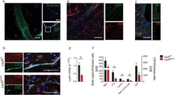
- Figure 1 Deletion of the leptin receptor (LepR) in brain endothelial and epithelial cells reduces leptin uptake by the brain . (A) Zs-Green reflecting expression of LepR was found in brain vessels (left and right panel) and in neurons of the VTA (right lower panel) of LepR-cre;Zs-Green mice. Nuclei are stained by DAPI (blue). Representative images are shown. Scale bar, 10 mum (left panel), 50 mum (right panels). (B and C) We used the reporter line HA-UPRT Fl to investigate in which cell types of the median eminence the Slco1c1-CreER T2 allele drives recombination. Immunofluorescent staining showed that HA-UPRT (red) was expressed in CD31-positive endothelial cells (green, B) of the cortex and mediobasal hypothalamus (C). There were no signs of recombination in vimentin-positive tanycytes (green, C). Scale bar, 50 mum. (D) Co-stainings of LepR (red) and CD31 (green) indicated that LepR was reduced in brain capillaries of Lepr beKO mice in comparison to LepR Fl controls. Capillaries in the cortex are depicted. (E) Relative LepR mRNA expression in primary brain endothelial cells. Values represent means +- SEM. Unpaired t test, *p < 0.05 (n = 3 mice/group). (F) Leptin uptake in the mediobasal hypothalamus (MBH), ventral tegmental area (VTA), cortex, cerebrospinal fluid (CSF), and the rest of the brain was reduced in LepR beKO mice compared to LepR Fl animals. Mice were perfused with 125 I-leptin for 30 min followed by a washout phase of 10 min. Values represent means +- SEM. Two-
- Submitted by
- Invitrogen Antibodies (provider)
- Main image

- Experimental details
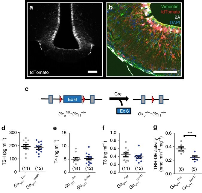
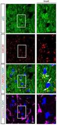
- Fig. 4 Basal activity of the HPT axis is not changed in mice with a tanycyte-specific deficiency of Galpha q/11. a tdTomato ( gray ) was expressed in tanycytes 2 weeks after injecting AAV-Dio2-iCre-2A-GFP in Ai14 reporter mice ( scale bar , 100 um). b Immunofluorescence staining of iCre-2A ( white ; 2A) and vimentin ( green ) showed colocalization in tdTomato-positive tanycytes ( red ; scale bar , 75 um). c Knockout strategy to generate Galpha q/11 tanKO mice. Cre recombinase was transduced by injecting AAV-Dio2-iCre-2A-GFP into the lateral ventricle of Galpha q fl/fl :: Galpha 11 -/- mice. AAV-Dio2-GFP injected Galpha q fl/fl :: Galpha 11 -/+ into the lateral ventricle were used as control ( Galpha q/11 Con ) d - f Basal plasma concentrations of TSH ( d ; p = 0.59), T4 ( e ; p = 0.75), and T3 ( f ; p = 0.34) 3 weeks after inducing the knockout by injecting rAAV ( Galpha q/11 Con , gray ; Galpha q/11 tanKO , blue ). g TRH-DE activity in Galpha q/11 Con mice ( gray ) and Galpha q/11 tanKO ( blue ) 3 weeks after rAAV injection. ** p = 0.0093 (two-tailed Student's t -test); mean +- S.E.M.; n , as indicated