Explore
 Validate
 Learn
Learn Western blot
 Immunohistochemistry
Antibody data
- Antibody Data
- Antigen structure
- References [2]
- Comments [0]
- Validations
- Immunohistochemistry [3]
Submit
Validation data
Reference
Comment
Report error
- Product number
- HPA001768 - Provider product page

- Provider
- Atlas Antibodies
- Proper citation
- Atlas Antibodies Cat#HPA001768, RRID:AB_1854252
- Product name
- Anti-MYO1C
- Antibody type
- Polyclonal
- Reactivity
- Human
- Host
- Rabbit
- Conjugate
- Unconjugated
- Antigen sequence
GEEETLRRLGLERNPQSYLYLVKGQCAKVSSINDK
SDWKVVRKALTVIDFTEDEVEDLLSIVASVLHLGN
IHFAANEESNAQVTTENQLKYLTRLLSVEGSTLRE
ALTHRKIIAKGEELLSPLNLEQAAYARDALAKAVY
SRTFTWLV- Isotype
- IgG
- Vial size
- 100 µl
- Storage
- Store at +4°C for short term storage. Long time storage is recommended at -20°C.
Submitted references Analysis of an independent tumor suppressor locus telomeric to Tp53 suggested Inpp5k and Myo1c as novel tumor suppressor gene candidates in this region.
The myosin motor Myo1c is required for VEGFR2 delivery to the cell surface and for angiogenic signaling.
Hedberg Oldfors C, Dios DG, Linder A, Visuttijai K, Samuelson E, Karlsson S, Nilsson S, Behboudi A
BMC genetics 2015 Jul 14;16:80
BMC genetics 2015 Jul 14;16:80
The myosin motor Myo1c is required for VEGFR2 delivery to the cell surface and for angiogenic signaling.
Tiwari A, Jung JJ, Inamdar SM, Nihalani D, Choudhury A
American journal of physiology. Heart and circulatory physiology 2013 Mar 1;304(5):H687-96
American journal of physiology. Heart and circulatory physiology 2013 Mar 1;304(5):H687-96
No comments: Submit comment
Enhanced validation
Supportive validation
- Submitted by
- Atlas Antibodies (provider)
- Enhanced method
- Orthogonal validation
- Main image

- Experimental details
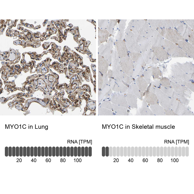
- Immunohistochemistry analysis in human lung and skeletal muscle tissues using Anti-MYO1C antibody. Corresponding MYO1C RNA-seq data are presented for the same tissues.
- Sample type
- HUMAN
Supportive validation
- Submitted by
- Atlas Antibodies (provider)
- Main image

- Experimental details
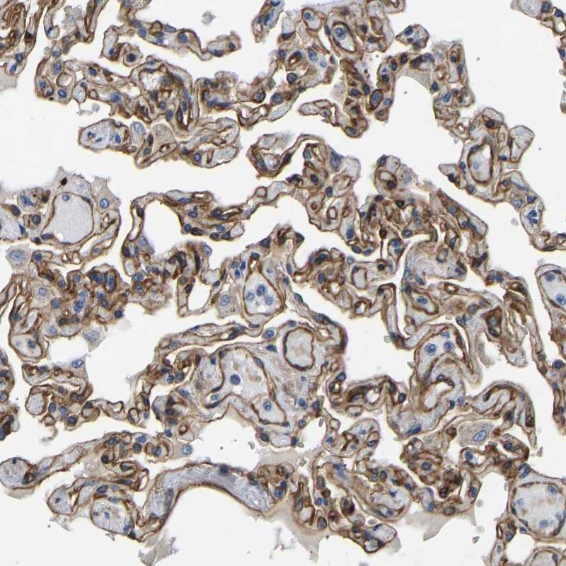
- Immunohistochemical staining of human lung shows high expression.
- Sample type
- HUMAN
- Submitted by
- Atlas Antibodies (provider)
- Main image
- Experimental details
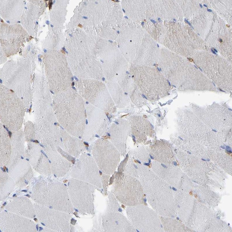
- Immunohistochemical staining of human skeletal muscle shows low expression as expected.
- Sample type
- HUMAN