Explore
 Validate
 Learn
Learn Western blot
Antibody data
- Antibody Data
- Antigen structure
- References [1]
- Comments [0]
- Validations
- Western blot [1]
- Immunocytochemistry [1]
- Immunohistochemistry [1]
Submit
Validation data
Reference
Comment
Report error
- Product number
- AF5896 - Provider product page

- Provider
- R&D Systems
- Product name
- Human/Mouse/Rat Semaphorin 5A Antibody
- Antibody type
- Polyclonal
- Description
- Immunogen affinity purified. Detects human, mouse, and rat Semaphorin 5A in direct ELISAs and Western blots. In direct ELISAs, less than 1% cross-reactivity with recombinant human (rh) Sema3B, rhSema4C, and rhSema6A is observed.
- Reactivity
- Human, Mouse, Rat
- Host
- Sheep
- Conjugate
- Unconjugated
- Antigen sequence
AAC09473- Isotype
- IgG
- Vial size
- 100 ug
- Concentration
- LYOPH
- Storage
- Use a manual defrost freezer and avoid repeated freeze-thaw cycles. 12 months from date of receipt, -20 to -70 °C as supplied. 1 month, 2 to 8 °C under sterile conditions after reconstitution. 6 months, -20 to -70 °C under sterile conditions after reconstitution.
Submitted references A Mutant p53-Dependent Embryonic Stem Cell Gene Signature Is Associated with Augmented Tumorigenesis of Stem Cells.
Koifman G, Shetzer Y, Eizenberger S, Solomon H, Rotkopf R, Molchadsky A, Lonetto G, Goldfinger N, Rotter V
Cancer research 2018 Oct 15;78(20):5833-5847
Cancer research 2018 Oct 15;78(20):5833-5847
No comments: Submit comment
Supportive validation
- Submitted by
- R&D Systems (provider)
- Main image
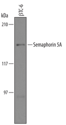
- Experimental details
- Detection of Mouse Semaphorin 5A by Western Blot. Western blot shows lysates of beta TC-6 mouse beta cell insulinoma cell line. PVDF Membrane was probed with 1 µg/mL of Sheep Anti-Human/Mouse/Rat Semaphorin 5A Antigen Affinity-purified Polyclonal Antibody (Catalog # AF5896) followed by HRP-conjugated Anti-Sheep IgG Secondary Antibody (Catalog # HAF016). A specific band was detected for Semaphorin 5A at approximately 135 kDa (as indicated). This experiment was conducted under reducing conditions and using Immunoblot Buffer Group 8.

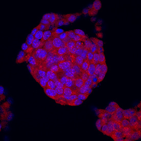
Supportive validation
- Submitted by
- R&D Systems (provider)
- Main image

- Experimental details
- Semaphorin 5A in beta TC-6 Mouse Cell Line. Semaphorin 5A was detected in immersion fixed beta TC-6 mouse beta cell insulinoma cell line using Sheep Anti-Human/Mouse/Rat Semaphorin 5A Antigen Affinity-purified Polyclonal Antibody (Catalog # AF5896) at 10 µg/mL for 3 hours at room temperature. Cells were stained using the NorthernLights™ 557-conjugated Anti-Sheep IgG Secondary Antibody (red; Catalog # NL010) and counterstained with DAPI (blue). Specific staining was localized to cytoplasm. View our protocol for Fluorescent ICC Staining of Cells on Coverslips.
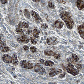
Supportive validation
- Submitted by
- R&D Systems (provider)
- Main image

- Experimental details
- Semaphorin 5A in Human Pancreas. Semaphorin 5A was detected in immersion fixed paraffin-embedded sections of human pancreas using Sheep Anti-Human/Mouse/Rat Semaphorin 5A Antigen Affinity-purified Polyclonal Antibody (Catalog # AF5896) at 5 µg/mL overnight at 4 °C. Tissue was stained using the Anti-Sheep HRP-DAB Cell & Tissue Staining Kit (brown; Catalog # CTS019) and counterstained with hemotoxylin (blue). Specific staining was localized to the plasma membranes of epithelial cells. View our protocol for Chromogenic IHC Staining of Paraffin-embedded Tissue Sections.