Explore
 Validate
 Learn
Learn Western blot
 ELISA
ELISAAntibody data
- Antibody Data
- Antigen structure
- References [0]
- Comments [0]
- Validations
- Western blot [2]
- Immunohistochemistry [3]
- Flow cytometry [6]
Submit
Validation data
Reference
Comment
Report error
- Product number
- NBP2-33108 - Provider product page

- Provider
- Novus Biologicals
- Product name
- Mouse Monoclonal Podocalyxin Like Antibody
- Antibody type
- Monoclonal
- Description
- Protein G purified. Podocalyxin is a member of the CD34 transmembrane sialomucin family. It is over-expressed on the podocyte foot projections and plays essential roles in kidney development and homeostasis, blood filtration and urine formation. It is also expressed on vascular endothelia, hematopoietic progenitors and a subset of neurons. Overexpression of podocalyxin may be linked to more aggressive tumor behavior. Podocalyxin antibody can identify podocytes in the urine (podocyturia) that may indicate glomerular disease, pre-eclampsia, and other kidney pathology.
- Reactivity
- Human, Mouse, Rat, Rabbit
- Host
- Mouse
- Isotype
- IgG
- Vial size
- 0.1 mg
- Concentration
- 1.0 mg/ml
- Storage
- Store at 4C short term. Aliquot and store at -20C long term. Avoid freeze-thaw cycles.
No comments: Submit comment
Supportive validation
- Submitted by
- Novus Biologicals (provider)
- Main image
- Experimental details
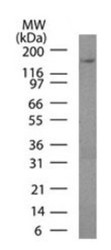
- Western Blot: Podocalyxin Like Antibody (3D3) - Azide and BSA Free [NBP2-33108] - HeLa lysate probed with Podocalyxin antibody at 3 ug/ml.

- Submitted by
- Novus Biologicals (provider)
- Main image
- Experimental details
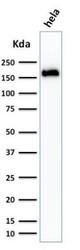
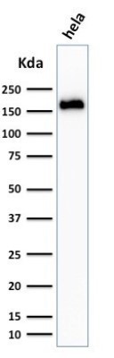
- Western Blot: Podocalyxin Like Antibody (3D3) - Azide and BSA Free [NBP2-33108] - Western Blot Analysis of HeLa cell lysate using Podocalyxin Like Antibody (3D3).
Supportive validation
- Submitted by
- Novus Biologicals (provider)
- Main image
- Experimental details
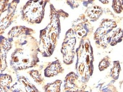
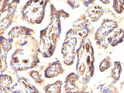
- Immunohistochemistry-Paraffin: Podocalyxin Like Antibody (3D3) - Azide and BSA Free [NBP2-33108] - Formalin-fixed, paraffin-embedded human Placenta stained with Podocalyxin Like Antibody (3D3).
- Submitted by
- Novus Biologicals (provider)
- Main image

- Experimental details
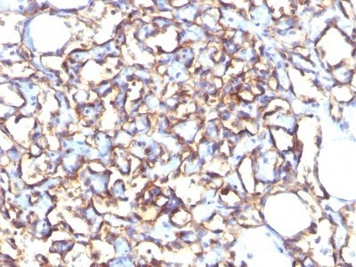
- Immunohistochemistry-Paraffin: Podocalyxin Like Antibody (3D3) - Azide and BSA Free [NBP2-33108] - Formalin-fixed, paraffin-embedded human Angiosarcoma stained with Podocalyxin Like Antibody (3D3).
- Submitted by
- Novus Biologicals (provider)
- Main image
- Experimental details
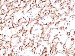
- Immunohistochemistry-Paraffin: Podocalyxin Like Antibody (3D3) - Azide and BSA Free [NBP2-33108] - Formalin-paraffin angiosarcoma stained with Podocalyxin Ab (3D3).

Supportive validation
- Submitted by
- Novus Biologicals (provider)
- Main image
- Experimental details
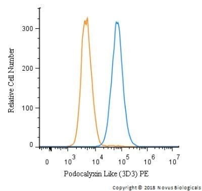
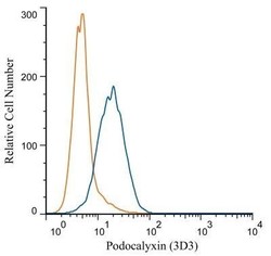
- Flow Cytometry: Podocalyxin Like Antibody (3D3) - Azide and BSA Free [NBP2-33108] - Analysis using PE conjugate of NBP2-25219. A cell surface stain was performed on HeLa cells with Podocalyxin antibody (3D3) NBP2-33108 (blue) and a matched isotype control NBP2-27287 (orange). Cells were incubated in an antibody dilution of 1:100 for 20 minutes at room temperature. Both antibodies were conjugated to PE.

- Submitted by
- Novus Biologicals (provider)
- Main image
- Experimental details
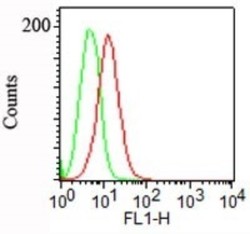
- Flow Cytometry: Podocalyxin Like Antibody (3D3) - Azide and BSA Free [NBP2-33108] - Surface staining of NCCIT cells using Podocalyxin antibody at 1 ug/10^6 cells. A FITC-conjugated anti mouse Ig secondary antibody was used along with this antibody .

- Submitted by
- Novus Biologicals (provider)
- Main image
- Experimental details
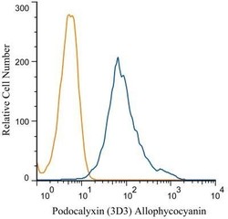
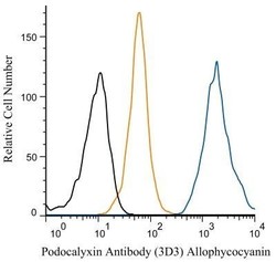
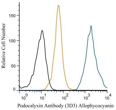
- Flow Cytometry: Podocalyxin Like Antibody (3D3) - Azide and BSA Free [NBP2-33108] - Analysis of Allophycocyanin conjugate of NBP2-25219. An intracellular stain was performed on HeLa cells with Podocalyxin (3D3) antibody NBP2-33108APC (blue) and a matched isotype control NBP2-27287APC (orange). Cells were fixed with 4% PFA and then permea
- Submitted by
- Novus Biologicals (provider)
- Main image
- Experimental details
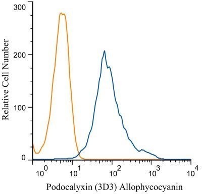
- Flow Cytometry: Podocalyxin Like Antibody (3D3) - Azide and BSA Free [NBP2-33108] - Analysis of Allophycocyanin conjugate of NBP2-25219. An intracellular stain was performed on SH-SY5Y cells with Podocalyxin (3D3) antibody NBP2-33108APC (blue) and a matched isotype control NBP2-27287APC (orange). Cells were fixed with 4% PFA and then per
- Submitted by
- Novus Biologicals (provider)
- Main image
- Experimental details
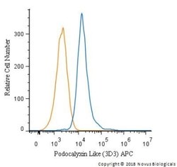
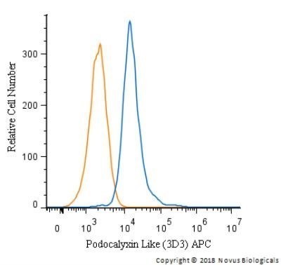
- Flow (Intracellular): Podocalyxin Like Antibody (3D3) - Azide and BSA Free [NBP2-33108] - An intracellular stain was performed on HeLa cells with Podocalyxin Like Antibody (3D3) NBP2-33108APC and a matched isotype control (orange). Cells were fixed with 4% PFA and then permeabilized with 0.1% saponin. Cells were incubated in an antibody dilution of 2.5 ug/mL for 30 minutes at room temperature. Both antibodies were conjugated to allophycocyanin.
- Submitted by
- Novus Biologicals (provider)
- Main image

- Experimental details
- Flow (Intracellular): Podocalyxin Like Antibody (3D3) - Azide and BSA Free [NBP2-33108] - An intracellular stain was performed on HeLa cells with Podocalyxin Like Antibody (3D3) NBP2-33108PE and a matched isotype control (orange). Cells were fixed with 4% PFA and then permeabilized with 0.1% saponin. Cells were incubated in an antibody dilution of 5 ug/mL for 30 minutes at room temperature. Both antibodies were conjugated to phycoerythrin. Image using the Azide and BSA Free form of this antibody.