Explore
 Validate
 Learn
Learn Western blot
 ELISA
ELISAAntibody data
- Antibody Data
- Antigen structure
- References [2]
- Comments [0]
- Validations
- Western blot [1]
- Immunocytochemistry [1]
- Immunohistochemistry [1]
- Flow cytometry [1]
Submit
Validation data
Reference
Comment
Report error
- Product number
- ABIN453850 - Provider product page

- Provider
- antibodies-online
- Product name
- anti-Thrombospondin 1 (THBS1) (N-Term) antibody
- Antibody type
- Polyclonal
- Antigen
- KLH conjugated synthetic peptide selected from the N-terminal region of human THBS1
- Description
- Affinity chromatography on Protein A
- Reactivity
- Human
- Host
- Rabbit
- Epitope
- N-Term
- Vial size
- 0.4 mL
- Concentration
- 0.25 mg/mL
- Storage
- Store the antibody undiluted at 2-8°C for one month or (in aliquots) at -20°C for longer.
- Handling
- Avoid repeated freezing and thawing.
Submitted references Structural characterization of the second TSP1-module of human thrombospondin.
C-mannosylation and O-fucosylation of the thrombospondin type 1 module.
Roszmusz E, Patthy A, Trexler M, Patthy L
Biochemical and biophysical research communications 2002 Aug 9;296(1):156-60
Biochemical and biophysical research communications 2002 Aug 9;296(1):156-60
C-mannosylation and O-fucosylation of the thrombospondin type 1 module.
Hofsteenge J, Huwiler KG, Macek B, Hess D, Lawler J, Mosher DF, Peter-Katalinic J
The Journal of biological chemistry 2001 Mar 2;276(9):6485-98
The Journal of biological chemistry 2001 Mar 2;276(9):6485-98
No comments: Submit comment
Supportive validation
- Submitted by
- antibodies-online (provider)
- Main image
- Experimental details
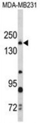
- Western blot analysis of THBS1 Antibody (N-term) Cat.-No AP18219PU-N in MDA-MB231 cell line lysates (35ug/lane). THBS1 (arrow) was detected using the purified Pab.

Supportive validation
- Submitted by
- antibodies-online (provider)
- Main image
- Experimental details
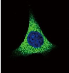
- Confocal immunofluorescent analysis of THBS1 Antibody (N-term) Cat.-No AP18219PU-N with MDA-MB231 cell followed by Alexa Fluor 488-conjugated goat anti-rabbit lgG (green). DAPI was used to stain the cell nuclear (blue).

Supportive validation
- Submitted by
- antibodies-online (provider)
- Main image

- Experimental details
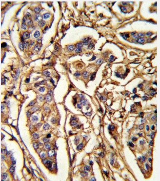
- Formalin-fixed and paraffin-embedded human breast carcinoma reacted with THBS1 Antibody (N-term) Cat.-No AP18219PU-N, which was peroxidase-conjugated to the secondary antibody, followed by DAB staining. This data demonstrates the use of this antibody for immunohistochemistry; clinical relevance has not been evaluated.
Supportive validation
- Submitted by
- antibodies-online (provider)
- Main image
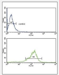
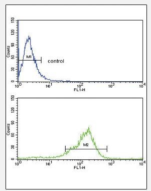
- Experimental details
- Flow cytometric analysis of MDA-MB231 cells using THBS1 Antibody (N-term) Cat.-No AP18219PU-N (bottom histogram) compared to a negative control cell (top histogram). FITC-conjugated goat-anti-rabbit secondary antibodies were used for the analysis