Explore
 Validate
 Learn
Learn Western blot
Antibody data
- Antibody Data
- Antigen structure
- References [0]
- Comments [0]
- Validations
- Western blot [8]
- Immunocytochemistry [1]
- Immunohistochemistry [1]
Submit
Validation data
Reference
Comment
Report error
- Product number
- PA1-16924 - Provider product page

- Provider
- Invitrogen Antibodies
- Product name
- Calreticulin Polyclonal Antibody
- Antibody type
- Polyclonal
- Antigen
- Other
- Description
- PA1-16924 is specific for the ~55 kDa Calreticulin protein.
- Concentration
- Conc. Not Determined
No comments: Submit comment
Supportive validation
- Submitted by
- Invitrogen Antibodies (provider)
- Main image

- Experimental details
- Detection of calreticulin in human kidney lysate using Product # PA1-16924 at 1:1,000. ECL exposure, 10 seconds.

- Submitted by
- Invitrogen Antibodies (provider)
- Main image
- Experimental details
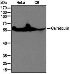
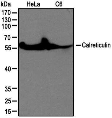
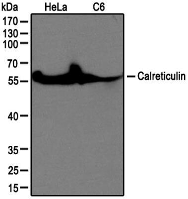
- Western blot analysis of Calreticulin in HeLa and C6 cell lysate. Samples were incubated in Calreticulin polyclonal antibody (Product # PA1-16924) using a dilution of 1:1000.
- Submitted by
- Invitrogen Antibodies (provider)
- Main image
- Experimental details
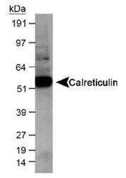
- Western blot analysis of Calreticulin in human kidney lysate. Samples were incubated in Calreticulin polyclonal antibody (Product # PA1-16924 using a dilution of 1:1,000. ECL exposure, 10 seconds.

- Submitted by
- Invitrogen Antibodies (provider)
- Main image
- Experimental details
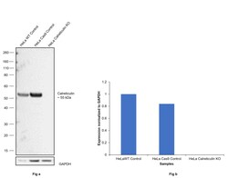
- Knockout of Calreticulin was achieved by CRISPR-Cas9 genome editing using LentiArray™ Lentiviral sgRNA (Product # A32042, Assay ID CRISPR1007602_LV) and LentiArray Cas9 Lentivirus (Product # A32064). Western blot analysis of Calreticulin was performed by loading 30 µg of HeLa wild type (Lane 1), HeLa Cas9 (Lane 2) and HeLa Calreticulin KO (Lane 3) whole cell extracts. The samples were electrophoresed using NuPAGE™ Novex™ 4-12% Bis-Tris Protein Gel (Product # NP0322BOX). Resolved proteins were then transferred onto a nitrocellulose membrane (Product # IB23001) by iBlot® 2 Dry Blotting System (Product # IB21001). The blot was probed with Calreticulin Polyclonal Antibody (Product # PA1-16924, 1:1000 dilution) and Goat anti-Rabbit IgG (H+L) Superclonal™ Recombinant Secondary Antibody, HRP (Product # A27036, 1:10,000 dilution) using the iBright™ FL1500 (Product # A44115). Chemiluminescent detection was performed usingSuperSignal™ West Dura Extended Duration Substrate (Product # 34076). Loss of signal upon CRISPR mediated knockout (KO) using the LentiArray™ CRISPR product line confirms that antibody is specific to Calreticulin.

- Submitted by
- Invitrogen Antibodies (provider)
- Main image

- Experimental details
- Western blot analysis of Calreticulin in HeLa and C6 cell lysate. Samples were incubated in Calreticulin polyclonal antibody (Product # PA1-16924) using a dilution of 1:1000.
- Submitted by
- Invitrogen Antibodies (provider)
- Main image

- Experimental details
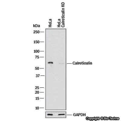
- Knockout validation by Western blot analysis of Calreticulin in lysates of HeLa human cervical epithelial carcinoma parental cell line and Calreticulin knockout (KO) HeLa cell line. Samples were incubated in Calreticulin polyclonal antibody (Product # PA1-16924) using a dilution of 1:2500 followed by a HRP-conjugated Anti-Rabbit IgG secondary antibody. Specific band was detected for Calreticulin at approximately 55 kDa (as indicated) in the parental HeLa cell line, but is not detectable in the knockout HeLa cell line. This experiment was conducted under reducing conditions.
- Submitted by
- Invitrogen Antibodies (provider)
- Main image

- Experimental details
- Western blot analysis of Calreticulin in 0.2 mg/mL HeLa lysate. Samples were incubated in Calreticulin polyclonal (Product # PA1-16924). This experiment was performed under reducing conditions using the 12-230 kDa separation system.

- Submitted by
- Invitrogen Antibodies (provider)
- Main image

- Experimental details
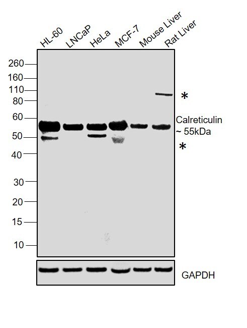
- Western blot was performed using Calreticulin Polyclonal Antibody (Product # PA1-16924) and a ~55kDa band corresponding to Calreticulin was observed across all the cell lines tested with a uncharacterized band (*). Whole cell extracts (30 µg lysate) of HL-60 (Lane 1), LNCaP(Lane 2), HeLa (Lane 3), MCF-7 (Lane 4), Mouse Liver (Lane 5) and Rat Liver (Lane 6) were electrophoresed using NuPAGE™ 4-12% Bis-Tris Protein Gel (Product # NP0321BOX). Resolved proteins were then transferred onto a Nitrocellulose membrane (Product # IB23001) by iBlot® 2 Dry Blotting System (Product # IB21001). The blot was probed with the primary antibody (1:1000 dilution) and detected by chemiluminescence with Goat anti-Rabbit IgG (H+L) Superclonal™ Recombinant Secondary Antibody, HRP (Product # A27036, 1:4000 dilution) using the iBright FL 1000 (Product # A32752). Chemiluminescent detection was performed using Novex® ECL Chemiluminescent Substrate Reagent Kit (Product # WP20005).
Supportive validation
- Submitted by
- Invitrogen Antibodies (provider)
- Main image

- Experimental details
- Immunocytochemistry analysis of Calreticulin in 3T3 cells. Samples were incubated in Calreticulin polyclonal antibody (Product # PA1-16924) followed by Alexa Fluor 488-conjugated Goat to rabbit IgG secondary antibody (green). Actin filaments were labeled with Alexa Fluor 568 phalloidin (red). DAPI was used to stain the cell nuclei (blue).

Supportive validation
- Submitted by
- Invitrogen Antibodies (provider)
- Main image
- Experimental details
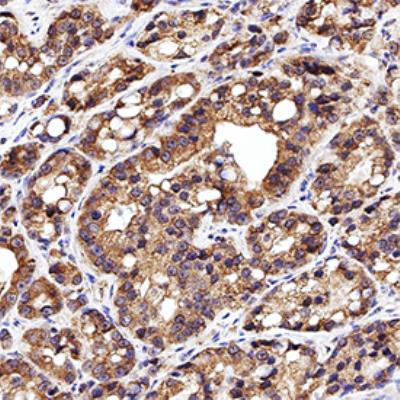
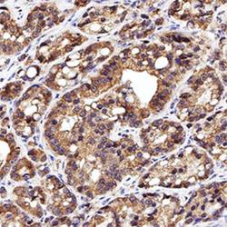
- Immunohistochemical analysis of Calreticulin in formalin-fixed paraffin-embedded human prostate cancer. Samples were incubated in Calreticulin polyclonal antibody (Product # PA1-16924) using a dilution of 1:20. Bond Rx autostainer (Leica Biosystems). The assay involved 20 minutes of heat induced antigen retrieval (HIER) using 10mM sodium citrate buffer (pH 6.0) and endogenous peroxidase quenching with peroxide block. The sections were incubated with primary antibody for 30 minutes and Bond Polymer Refine Detection (Leica Biosystems) with DAB was used for signal development followed by counterstaining with hematoxylin. Whole slide scanning and capturing of representative images was performed using Aperio AT2 (Leica Biosystems). Cytoplasmic staining in glands was observed. Staining was performed by Histowiz.