Explore
 Validate
 Learn
Learn Western blot
 ELISA
ELISAAntibody data
- Antibody Data
- Antigen structure
- References [0]
- Comments [0]
- Validations
- Western blot [9]
- Immunocytochemistry [2]
- Immunohistochemistry [1]
- Flow cytometry [1]
- Chromatin Immunoprecipitation [1]
Submit
Validation data
Reference
Comment
Report error
- Product number
- MA5-17169 - Provider product page

- Provider
- Invitrogen Antibodies
- Product name
- RUNX3 Monoclonal Antibody (2B3)
- Antibody type
- Monoclonal
- Antigen
- Purifed from natural sources
- Description
- MA5-17169 targets RUNX3 in indirect ELISA, FACS, ICC, IHC, IF and WB applications and shows reactivity with Human and Mouse samples. The MA5-17169 immunogen is purified recombinant fragment of human RUNX3 (amino acids:186-252) expressed in E. Coli. MA5-17169 detects RUNX3 which has a predicted molecular weight of approximately 44.4kDa.
- Reactivity
- Human, Mouse
- Host
- Mouse
- Isotype
- IgG
- Antibody clone number
- 2B3
- Vial size
- 100 µg
- Concentration
- 1 mg/mL
- Storage
- Store at 4°C short term. For long term storage, store at -20°C, avoiding freeze/thaw cycles.
No comments: Submit comment
Supportive validation
- Submitted by
- Invitrogen Antibodies (provider)
- Main image

- Experimental details
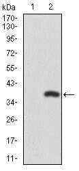
- Western blot analysis of RUNX3 using RUNX3 monoclonal antibody (Product # MA5-17169) in HEK293 (1) and RUNX3 (AA: 186-252) human IgG Fc transfected HEK293 (2) cell lysate.
- Submitted by
- Invitrogen Antibodies (provider)
- Main image

- Experimental details
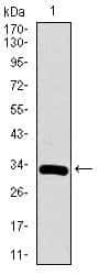
- Western blot analysis of RUNX3 using a RUNX3 monoclonal antibody (Product # MA5-17169) against a human RUNX3 recombinant protein.
- Submitted by
- Invitrogen Antibodies (provider)
- Main image
- Experimental details
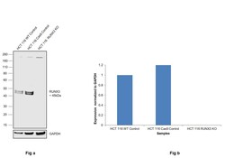
- Knockout of RUNX3 was achieved by CRISPR-Cas9 genome editing using LentiArray™ Lentiviral sgRNA (Product # A32042, Assay ID ) and LentiArray Cas9 Lentivirus (Product # A32064). Western blot analysis of RUNX3 was performed by loading 60 µg of HCT 116 wild type (Lane 1), HCT 116 Cas9 (Lane 2) andHCT 116 RUNX3 KO (Lane 3) whole cell extracts. The samples were electrophoresed using NuPAGE™ Novex™ 4-12% Bis-Tris Protein Gel (Product # NP0321BOX). Resolved proteins were then transferred onto a nitrocellulose membrane (Product # IB23001) by iBlot® 2 Dry Blotting System (Product # IB21001). The blot was probed with Anti-RUNX3 Monoclonal Antibody (2B3) (Product # MA5-17169, 1:500 dilution) and Goat anti-Mouse IgG (H+L) Superclonal™ Recombinant Secondary Antibody, HRP (Product # A28177, 1:6000 dilution) using the iBright FL 1000 (Product # A32752). Chemiluminescent detection was performed using SuperSignal™ West Dura Extended Duration Substrate (Product # 34076). Loss of signal upon CRISPR mediated knockout (KO) using the LentiArray™ CRISPR product line confirms that antibody is specific to RUNX3. An uncharacterized band observed around ~240 kDa in all the samples.

- Submitted by
- Invitrogen Antibodies (provider)
- Main image

- Experimental details
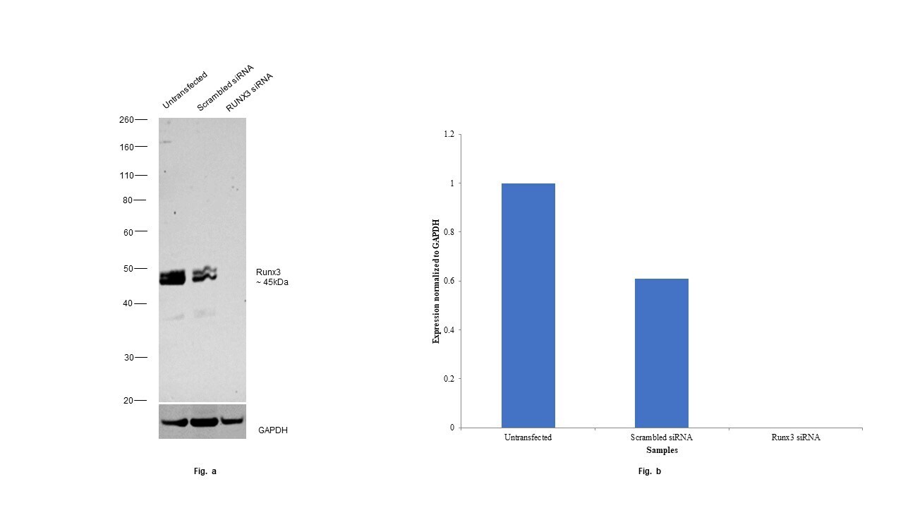
- Knockdown of RUNX3 was achieved by transfecting HCT 116 with RUNX3 specific siRNAs (Silencer® select Product # S2467, S2468). Western blot analysis (Fig. a) was performed using whole cell extracts from the RUNX3 knockdown cells (lane 3), non-targeting scrambled siRNA transfected cells (lane 2) and untransfected cells (lane 1). The blot was probed with RUNX3 Monoclonal Antibody (2B3) (Product # MA5-17169, 1:1500) and Goat anti-Mouse IgG (H+L) Superclonal™ Recombinant Secondary Antibody, HRP (Product # A28177, 1:20,000). Densitometric analysis of this western blot is shown in histogram (Fig. b). Decrease in signal upon siRNA mediated knock down confirms that antibody is specific to RUNX3.
- Submitted by
- Invitrogen Antibodies (provider)
- Main image
- Experimental details
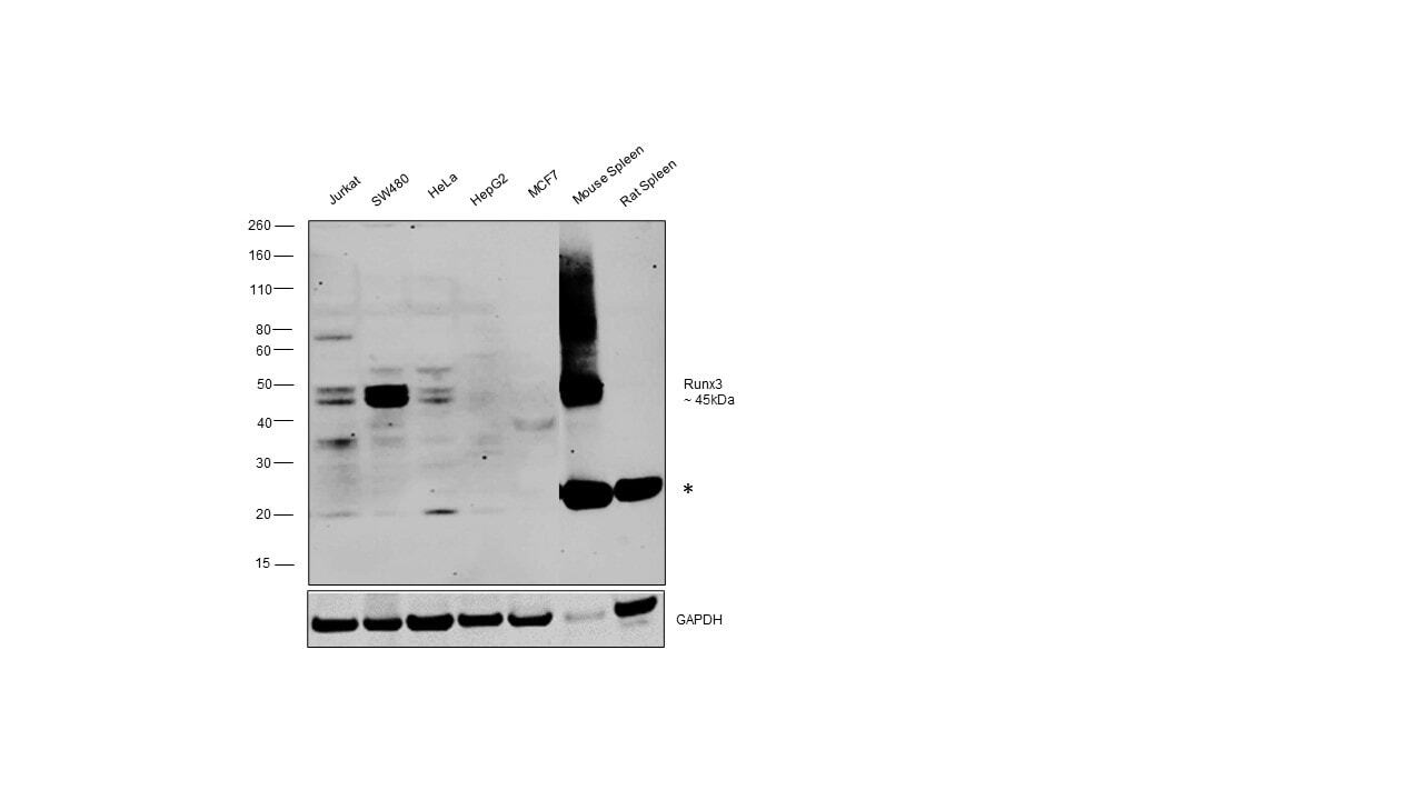
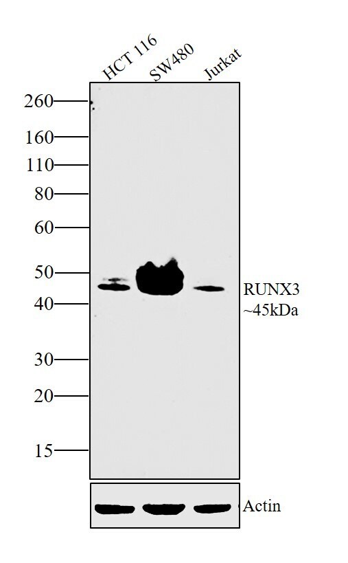
- Western blot was performed using RUNX3 Monoclonal Antibody (2B3) (Product # MA5-17169) and a ~45 kDa band corresponding to RUNX3 was observed only in Jurkat, SW480, HeLa and Mouse Spleen. Whole cell extracts (30 µg lysate) of Jurkat (Lane 1), SW480 (Lane 2), HeLa (Lane 3), Hep G2 (Lane 4), MCF7 (Lane 5), Mouse Spleen (Lane 6), Rat Spleen (Lane 7) were electrophoresed using NuPAGE™ 4-12% Bis-Tris Protein Gel (Product # NP0322BOX), 12 well. Resolved proteins were then transferred onto a nitrocellulose membrane (Product # IB23001) by iBlot® 2 Dry Blotting System (Product # IB21001). The blot was probed with the primary antibody (1:1500) and detected by chemiluminescence with Goat anti-Mouse IgG (H+L) Superclonal™ Recombinant Secondary Antibody, HRP (Product # A28177, 1:20,000) using the iBright™ FL1500 Imaging System (Product # A44115). Chemiluminescent detection was performed using SuperSignal™ West Pico PLUS Chemiluminescent Substrate (Product # 34580).
- Submitted by
- Invitrogen Antibodies (provider)
- Main image

- Experimental details
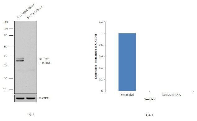
- Knockdown of RUNX3 was achieved by transfecting HCT 116 cells with RUNX3 specific siRNAs (Silencer® select Product # s2467, s2468). Western blot analysis (Fig. a) was performed using whole cell extracts from the RUNX3 knockdown cells (lane 2) and non-specific scrambled siRNA transfected cells (lane 1). The blots were probed with RUNX3 Monoclonal Antibody (Product # MA5-17169, 1:1000 dilution) and Goat anti-Mouse IgG (H+L) Superclonal™ Secondary Antibody, HRP conjugate (Product # A28177, 0.25 µg/mL, 1:4000 dilution). Densitometric analysis of this western blot is shown in histogram (Fig. b). Decrease in signal upon siRNA mediated knock down confirms that antibody is specific to RUNX3.
- Submitted by
- Invitrogen Antibodies (provider)
- Main image

- Experimental details
- Western blot analysis was performed on modified whole cell extracts (1% SDS) of HCT 116 (Lane 1), SW480 (Lane 2) and Jurkat (Lane 3). The blot was probed with RUNX3 Monoclonal Antibody (2B3) (Product # MA5-17169, 1:1000 dilution) and detected by chemiluminescence using Goat anti-Mouse IgG (H+L) Superclonal™ Secondary Antibody, HRP conjugate (Product # A28177, 0.25 µg/mL, 1:4000 dilution). A 45 kDa band corresponding to RUNX3 was observed across the panel.
- Submitted by
- Invitrogen Antibodies (provider)
- Main image

- Experimental details
- Western blot analysis of RUNX3 using a RUNX3 monoclonal antibody (Product # MA5-17169) against a human RUNX3 recombinant protein.

- Submitted by
- Invitrogen Antibodies (provider)
- Main image

- Experimental details
- Western blot analysis of RUNX3 using RUNX3 monoclonal antibody (Product # MA5-17169) in HEK293 (1) and RUNX3 (AA: 186-252) human IgG Fc transfected HEK293 (2) cell lysate.

Supportive validation
- Submitted by
- Invitrogen Antibodies (provider)
- Main image

- Experimental details
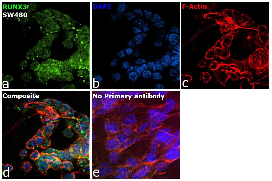
- Immunofluorescence analysis of RUNX3 was performed using 70% confluent log phase SW480 cells. The cells were fixed with 4% paraformaldehyde for 10 minutes, permeabilized with 0.1% Triton™ X-100 for 10 minutes, and blocked with 2% BSA for 45 minutes at room temperature. The cells were labeled with RUNX3 Monoclonal Antibody (2B3) (Product # MA5-17169) at 1:100 in 0.1% BSA, incubated at 4 degree celsius overnight and then labeled with Donkey anti-Mouse IgG (H+L) Highly Cross-Adsorbed Secondary Antibody, Alexa Fluor™ Plus 488 (Product # A32766, 1:2000), for 45 minutes at room temperature (Panel a: Green). Nuclei (Panel b:Blue) were stained with ProLong™ Diamond Antifade Mountant with DAPI (Product # P36962). F-actin (Panel c: Red) was stained with Rhodamine Phalloidin (Product # R415, 1:300). Panel d represents the merged image showing nucleus and cytoplasmic localization. Panel e represents control cells with no primary antibody to assess background. The images were captured at 60X magnification.
- Submitted by
- Invitrogen Antibodies (provider)
- Main image
- Experimental details
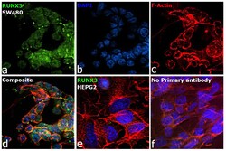
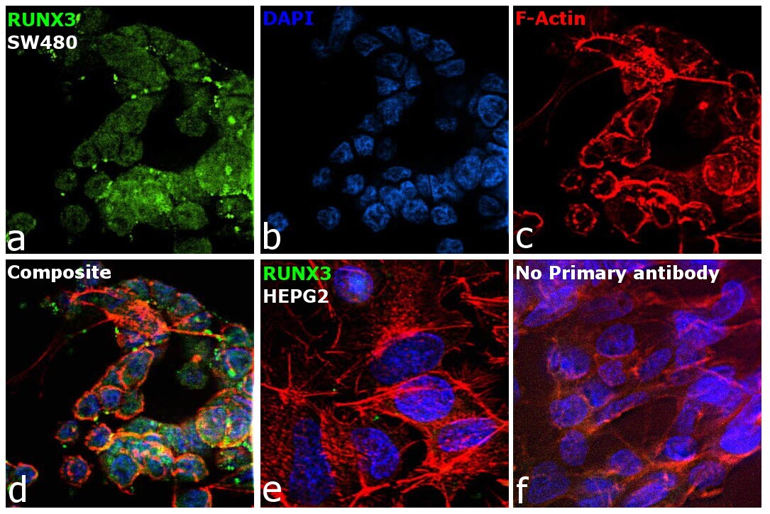
- Immunofluorescence analysis of RUNX3 was performed using 70% confluent log phase SW480 and Hep G2 cells. The cells were fixed with 4% paraformaldehyde for 10 minutes, permeabilized with 0.1% Triton™ X-100 for 10 minutes, and blocked with 2% BSA for 45 minutes at room temperature. The cells were labeled with RUNX3 Monoclonal Antibody (2B3) (Product # MA5-17169) at 1:100 in 0.1% BSA, incubated at 4 degree celsius overnight and then labeled with Donkey anti-Mouse IgG (H+L) Highly Cross-Adsorbed Secondary Antibody, Alexa Fluor™ Plus 488 (Product # A32766, 1:2000), for 45 minutes at room temperature (Panel a: Green). Nuclei (Panel b: Blue) were stained with ProLong™ Diamond Antifade Mountant with DAPI (Product # P36962). F-actin (Panel c: Red) was stained with Rhodamine Phalloidin (Product # R415, 1:300). Panel d represents the merged image showing nucleus and cytoplasmic localization. Panel e represents Hep G2 showing no signal. Panel f represents control cells with no primary antibody to assess background. The images were captured at 60X magnification.
Supportive validation
- Submitted by
- Invitrogen Antibodies (provider)
- Main image
- Experimental details
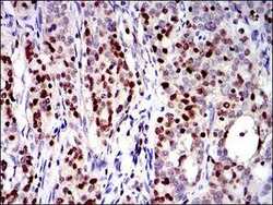
- Immunohistochemical analysis of paraffin-embedded cervical cancer tissues using RUNX3 monoclonal antibody (Product # MA5-17169) followed with DAB staining.

Supportive validation
- Submitted by
- Invitrogen Antibodies (provider)
- Main image
- Experimental details
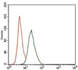
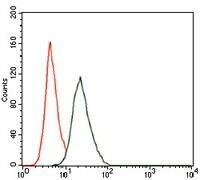
- Flow cytometric analysis of NIH3T3 cells using RUNX3 monoclonal antibody (Product # MA5-17169) (green) and negative control (red).
Supportive validation
- Submitted by
- Invitrogen Antibodies (provider)
- Main image
- Experimental details
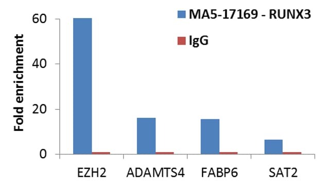
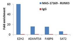
- Enrichment of endogenous RUNX3 protein at specific gene loci using Anti-RUNX3 Antibody: Chromatin Immunoprecipitation (ChIP) was performed using Anti-RUNX3 Mouse Monoclonal antibody (Product # MA5-17169, 5 µg) on sheared chromatin from 2 million HCT 116 cells using the MAGnify ChIP System (Product # 49-2024). Normal Rabbit IgG was used as a negative IP control. The purified DNA was analyzed by qPCR with PCR primer pairs over the promoters of EZH2, ADAMTS4 and FABP6 (positive) and SAT2 satellite repeats (negative). Data is presented as fold enrichment of the antibody signal versus the negative control IgG using the comparative CT method.