Explore
 Validate
 Learn
Learn Western blot
Antibody data
- Antibody Data
- Antigen structure
- References [1]
- Comments [0]
- Validations
- Western blot [4]
- Immunocytochemistry [1]
- Immunohistochemistry [2]
- Other assay [3]
Submit
Validation data
Reference
Comment
Report error
- Product number
- PA5-27210 - Provider product page

- Provider
- Invitrogen Antibodies
- Product name
- MMP1 Polyclonal Antibody
- Antibody type
- Polyclonal
- Antigen
- Recombinant protein fragment
- Description
- Recommended positive controls: Raji, HUVEC, dermal fibroblast cells. Predicted reactivity: Pig (90%), Rabbit (86%), Bovine (83%). Store product as a concentrated solution. Centrifuge briefly prior to opening the vial.
- Reactivity
- Human, Mouse
- Host
- Rabbit
- Isotype
- IgG
- Vial size
- 100 µL
- Concentration
- 0.54 mg/mL
- Storage
- Store at 4°C short term. For long term storage, store at -20°C, avoiding freeze/thaw cycles.
Submitted references ITLN1 modulates invasive potential and metabolic reprogramming of ovarian cancer cells in omental microenvironment.
Au-Yeung CL, Yeung TL, Achreja A, Zhao H, Yip KP, Kwan SY, Onstad M, Sheng J, Zhu Y, Baluya DL, Co NN, Rynne-Vidal A, Schmandt R, Anderson ML, Lu KH, Wong STC, Nagrath D, Mok SC
Nature communications 2020 Jul 15;11(1):3546
Nature communications 2020 Jul 15;11(1):3546
No comments: Submit comment
Supportive validation
- Submitted by
- Invitrogen Antibodies (provider)
- Main image

- Experimental details
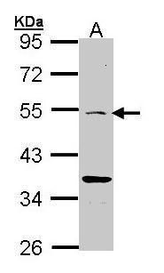
- Western blot analysis of MMP1 using 30 µg of Raji lysate. Samples were loaded onto a 10% SDS-PAGE gel and probed with a MMP1 polyclonal antibody (Product # PA5-27210) at a dilution of 1:1000.
- Submitted by
- Invitrogen Antibodies (provider)
- Main image

- Experimental details
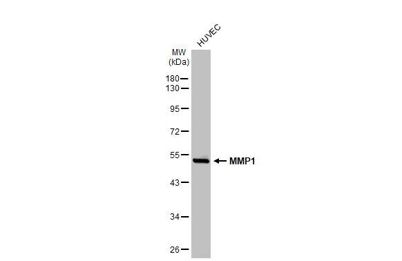
- Western blot analysis of MMP1 was performed by separating 30 µg of whole cell extract by 10% SDS-PAGE. Proteins were transferred to a membrane and probed with a MMP1 Polyclonal Antibody (Product # PA5-27210) at a dilution of 1:2500. The HRP-conjugated anti-rabbit IgG antibody was used to detect the primary antibody.

- Submitted by
- Invitrogen Antibodies (provider)
- Main image

- Experimental details
- Western Blot using MMP1 Polyclonal Antibody (Product # PA5-27210). Whole cell extract (30 µg) was separated by 10% SDS-PAGE, and the membrane was blotted with MMP1 Polyclonal Antibody (Product # PA5-27210) diluted at 1:2,500. The HRP-conjugated anti-rabbit IgG antibody was used to detect the primary antibody.
- Submitted by
- Invitrogen Antibodies (provider)
- Main image
- Experimental details
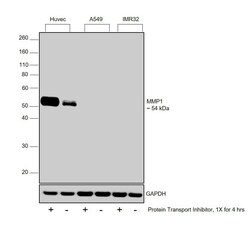
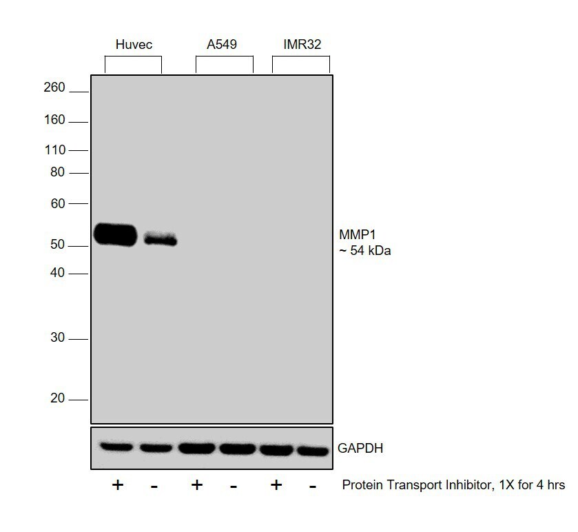
- Western blot was performed using Anti-MMP1 Polyclonal Antibody (Product # PA5-27210) and a 54 kDa band corresponding to MMP1 was observed in HUVEC, the positive model for MMP1 and increased upon Protein Transport Inhibitor (PTI) treatment while not detected in A549 or IMR32 both of which are reported to be low for MMP1, relative to HUVEC. Whole cell extracts (30 µg lysate) of Huvec treated with PTI (1X for 4hrs) (Lane 1), Huvec untreated (Lane 2), A549 treated with PTI (Lane 3), A549 untreated (Lane 4), IMR32 treated with PTI (Lane 5) and IMR32 untreated (Lane 6) were electrophoresed using NuPAGE™ 10% Bis-Tris Protein Gel (Product # NP0302BOX). Resolved proteins were then transferred onto a nitrocellulose membrane (Product # IB23001) by iBlot® 2 Dry Blotting System (Product # IB21001). The blot was probed with the primary antibody (0.5µg/ml) and detected by chemiluminescence with Goat anti-Rabbit IgG (H+L), Superclonal™ Recombinant Secondary Antibody, HRP (Product # A27036, 1:4000 dilution) using the iBright FL 1000 (Product # A32752). Chemiluminescent detection was performed using Novex® ECL Chemiluminescent Substrate Reagent Kit (Product # WP20005)..
Supportive validation
- Submitted by
- Invitrogen Antibodies (provider)
- Main image
- Experimental details
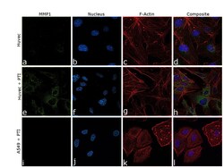
- Immunofluorescence analysis of MMP1 was performed using 70% confluent log phase HUVEC and A549 untreated and Protein Transport Inhibitor (PTI) treated cells. The cells were fixed with 4% Paraformaldehyde for 10 minutes, permeabilized with 0.1% Triton™ X-100 for 10 minutes, and blocked with 2% BSA for 10 minutes at room temperature. The cells were labeled with MMP1 Polyclonal Antibody (Product # PA5-27210) at 5 µg/mL in 0.1% BSA, incubated at 4 degree celsius overnight and then labeled with Goat anti-Rabbit IgG (H+L), Superclonal™ Recombinant Secondary Antibody, Alexa Fluor 488 (Product # A27034) at 1:2000 dilution for 45 minutes at room temperature (Panel a, e, i: Green). Nuclei (Panel b, f, j: Blue) were stained with SlowFade® Gold Antifade Mountant with DAPI (Product # S36938). F-actin (Panel c, g, k: Red) was stained with Rhodamine Phalloidin (Product # R415, 1:300). Panel d, h, l represents the merged image showing increased cytoplasmic localization of MMP1 in Huvec upon PTI treatment in comparison to untreated Huvec and also A549 treated with PTI which is reported as a low expressing model for A549 (Untreated A549 data not shown). The images were captured at 60X magnification.

Supportive validation
- Submitted by
- Invitrogen Antibodies (provider)
- Main image
- Experimental details
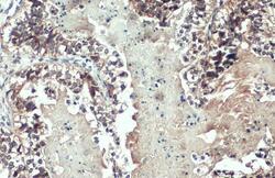
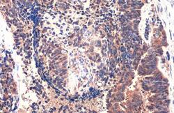
- MMP1 Polyclonal Antibody detects MMP1 protein at cytoplasm by immunohistochemical analysis. Sample: Paraffin-embedded human endometrial carcinoma. MMP1 stained by MMP1 Polyclonal Antibody (Product # PA5-27210) diluted at 1:500. Antigen Retrieval: Citrate buffer, pH 6.0, 15 min.
- Submitted by
- Invitrogen Antibodies (provider)
- Main image
- Experimental details
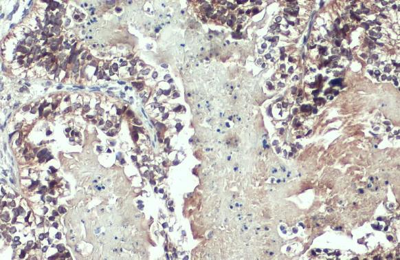
- MMP1 Polyclonal Antibody detects MMP1 protein at cytoplasm by immunohistochemical analysis. Sample: Paraffin-embedded human endometrial carcinoma. MMP1 stained by MMP1 Polyclonal Antibody (Product # PA5-27210) diluted at 1:500. Antigen Retrieval: Citrate buffer, pH 6.0, 15 min.

Supportive validation
- Submitted by
- Invitrogen Antibodies (provider)
- Main image

- Experimental details
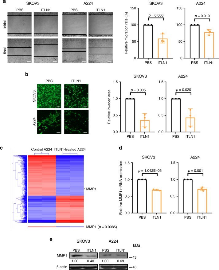
- Fig. 3 MMP1 mediates the effect of ITLN1 on suppressing ovarian cancers' motility. Representative microscopic images of ( a ) a wound-healing assay show that ITLN1 suppressed cell migration ability in SKOV3 and A224; b a cell invasion assay show that ITLN1 suppressed cell invasive potential in SKOV3 and A224. Bar = 50 mum. Results in the bar charts, presented as mean +- SD (two-tailed t -test), show the average from three independent experiments with duplicated samples. PBS phosphate-buffered saline. c Heat map from a transcriptome profiling analysis shows that MMP1 expression is significantly decreased in ITLN1-treated A224 ( n = 2) compared with control A224 without ITLN1 treatment ( n = 2). d QRT-PCR analysis shows a lower MMP1 mRNA level in ITLN1-treated SKOV3 and A224 compared with control cells treated with PBS. Results, as presented as mean +- SD (two-tailed t -test), show the average from three independent experiments. e Western blot analysis shows a lower MMP1 protein level in ITLN1-treated SKOV3 and A224 compared with control cells treated with PBS. beta-actin served as a loading control. Relative normalized protein levels with respect to the corresponding control are presented. Three independent experiments were performed.
- Submitted by
- Invitrogen Antibodies (provider)
- Main image
- Experimental details
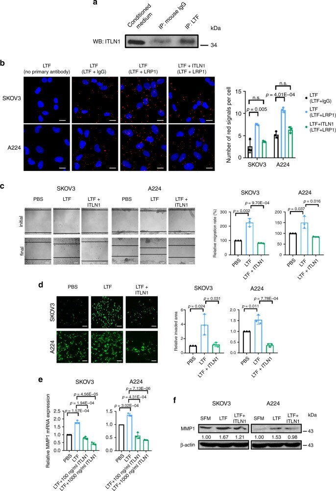
- Fig. 4 ITLN1 abrogates LTF's effects on ovarian cancer cells' motility. a Western blot analysis shows ITLN1 on proteins pulled down by an anti-LTF antibody. Normal mouse IgG served as a control. ITLN1 band size = 34 kDa. b Representative microscopic images from a Duolink proximity ligation assay (PLA) on SKOV3 and A224 shows the interaction between LTF and LRP1 while ITLN1 reduced the interaction. Red fluorescent signals indicate protein-protein interaction; nuclei were stained with 4,6-diamidino-2-phenylindole (DAPI) blue. Staining with no primary antibody and with anti-LTF antibody plus normal rabbit IgG served as controls. Bar = 5 mum. Representative microscopic images show that LTF induced c cell migration ability and d cell invasion potential in SKOV3 and A224 compared with control cells, while ITLN1 counteracted the effect. Bar = 50 mum. e Bar charts show that LTF upregulated the relative MMP1 mRNA expression in SKOV3 and A224, while ITLN1 counteracted the effect. f Western blot analysis shows that LTF upregulated MMP1 protein expression in SKOV3 and A224, while ITLN1 counteracted the effect. beta-actin served as a loading control. Relative normalized protein levels with respect to the corresponding control are presented. Three independent experiments were performed. b , e Results from three independent experiments were averaged and are shown as mean +- SD (two-tailed t -test). n. s. not significant ( p > 0.05).
- Submitted by
- Invitrogen Antibodies (provider)
- Main image
- Experimental details
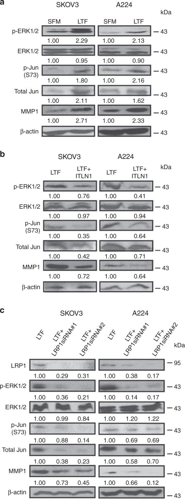
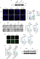
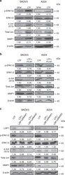
- Fig. 5 ITLN1 inhibits the LTF/LRP1/MMP1 signaling pathway. Western blot analyses show ( a ) higher protein levels of p-ERK1/2, p-Jun (S73), total Jun, and MMP1 in LTF-treated (100 mug mL -1 ) SKOV3 and A224 compared with control cells without treatment; b lower protein levels of p-ERK1/2, p-Jun (S73), total Jun, and MMP1 in LTF-treated SKOV3 and A224 with ITLN1 (500 ng mL -1 ) compared with control cells without ITLN1; and c lower protein levels of LRP1, p-ERK1/2, p-Jun (S73), total Jun, and MMP1 in LTF-treated SKOV3 and A224 with LRP1-specific siRNAs compared with control cells with negative siRNA. beta-actin served as a loading control. Relative normalized protein levels with respect to the corresponding control are presented. Three independent experiments were performed.