 Explore
 Validate
 Learn
Learn Immunohistochemistry
Antibody data
- Antibody Data
- Antigen structure
- References [1]
- Comments [0]
- Validations
- Immunohistochemistry [15]
Submit
Validation data
Reference
Comment
Report error
- Product number
- HPA020339 - Provider product page

- Provider
- Atlas Antibodies
- Proper citation
- Atlas Antibodies Cat#HPA020339, RRID:AB_1845131
- Product name
- Anti-ATXN2
- Antibody type
- Polyclonal
- Reactivity
- Human
- Host
- Rabbit
- Conjugate
- Unconjugated
- Antigen sequence
EGHSINTRENKYIPPGQRNREVISWGSGRQNSPRM
GQPGSGSMPSRSTSHTSDFNPNSGSDQRVVNGGVP
WPSPCPSPSSRPPSRYQSGPNSLPPRAATPTRPPS
RPPSRPSRPPSHPSAHGSPAPVSTMPKRMSSE- Isotype
- IgG
- Vial size
- 100 µl
- Storage
- Store at +4°C for short term storage. Long time storage is recommended at -20°C.
Submitted references Ataxin-2-like is a regulator of stress granules and processing bodies.
Kaehler C, Isensee J, Nonhoff U, Terrey M, Hucho T, Lehrach H, Krobitsch S
PloS one 2012;7(11):e50134
PloS one 2012;7(11):e50134
No comments: Submit comment
Enhanced validation
Enhanced validation
Supportive validation
- Submitted by
- Atlas Antibodies (provider)
- Enhanced method
- Orthogonal validation
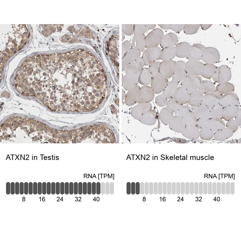
- Main image

- Experimental details
- Immunohistochemistry analysis in human testis and skeletal muscle tissues using HPA020339 antibody. Corresponding ATXN2 RNA-seq data are presented for the same tissues.
- Sample type
- HUMAN
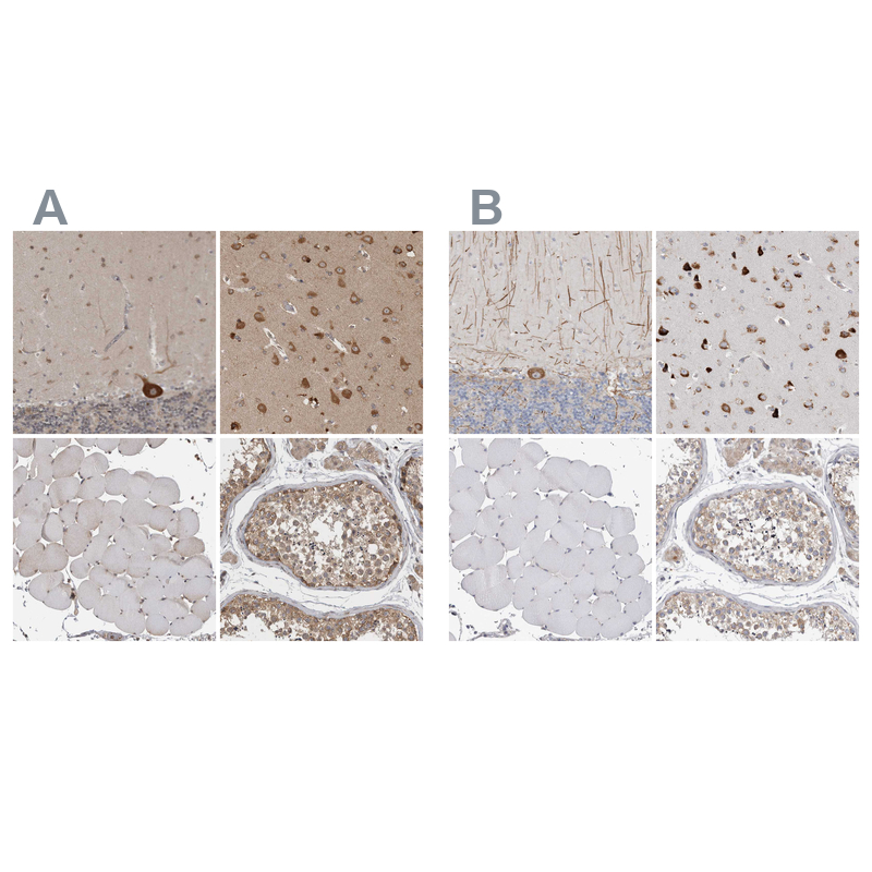
Enhanced validation
- Submitted by
- Atlas Antibodies (provider)
- Enhanced method
- Independent antibody validation
- Main image

- Experimental details
- Immunohistochemical staining of human cerebellum, cerebral cortex, skeletal muscle and testis using Anti-ATXN2 antibody HPA020339 (A) shows similar protein distribution across tissues to independent antibody HPA018295 (B).
Supportive validation
- Submitted by
- Atlas Antibodies (provider)
- Main image

- Experimental details
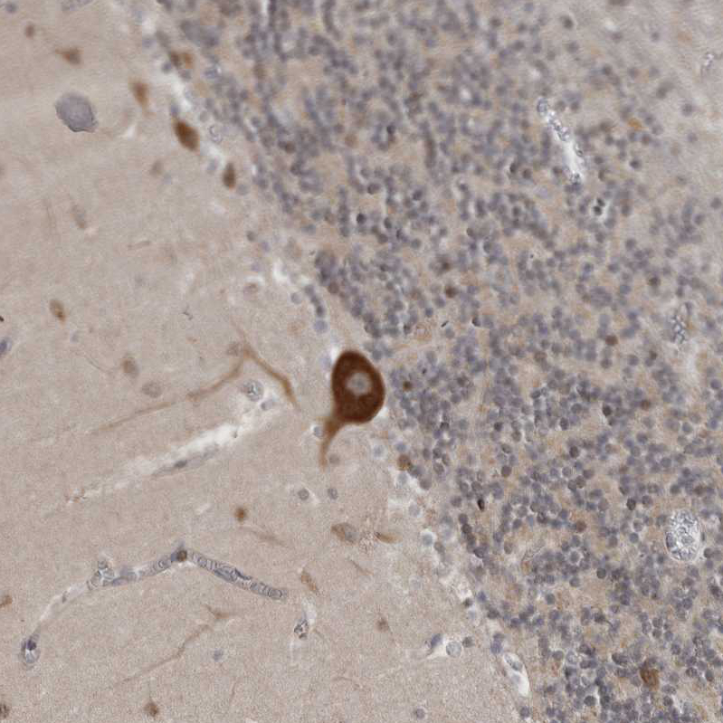
- Immunohistochemical staining of human cerebral cortex shows strong cytoplasmic positivity in neurons.

- Submitted by
- Atlas Antibodies (provider)
- Main image
- Experimental details
- Immunohistochemical staining of human testis shows high expression.
- Sample type
- HUMAN
- Submitted by
- Atlas Antibodies (provider)
- Main image

- Experimental details
- Immunohistochemical staining of human skeletal muscle shows low expression as expected.
- Sample type
- HUMAN
- Submitted by
- Atlas Antibodies (provider)
- Main image

- Experimental details
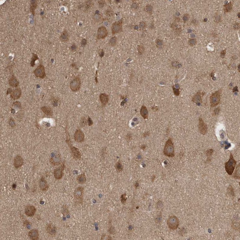
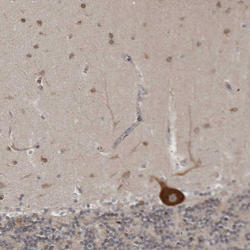
- Immunohistochemical staining of human cerebral cortex shows moderate to strong cytoplasmic positivity in neurons.
- Sample type
- HUMAN
- Submitted by
- Atlas Antibodies (provider)
- Main image

- Experimental details
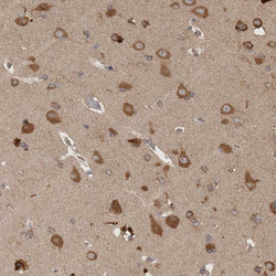
- Immunohistochemical staining of human cerebellum shows moderate to strong cytoplasmic positivity in purkinje cells.
- Sample type
- HUMAN
- Submitted by
- Atlas Antibodies (provider)
- Main image

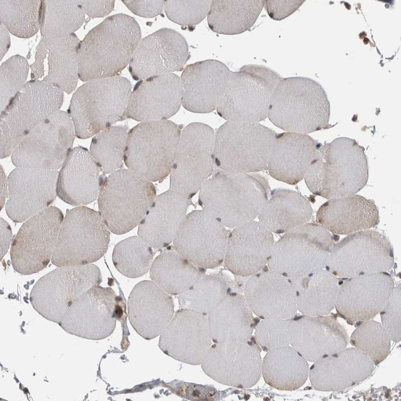
- Experimental details
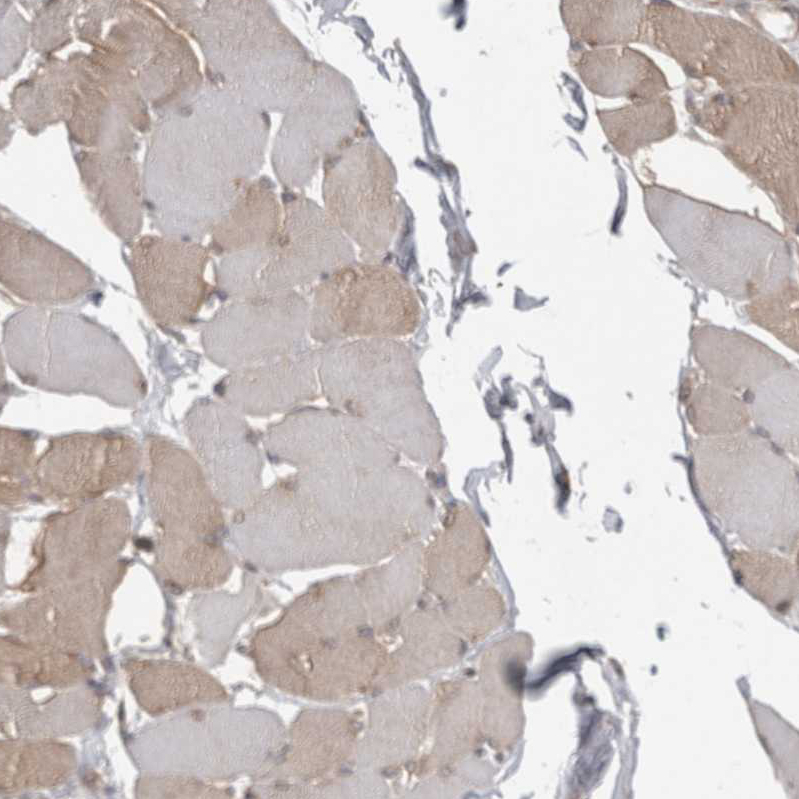
- Immunohistochemical staining of human skeletal muscle shows weak to moderate cytoplasmic positivity in a subset of myocytes.
- Sample type
- HUMAN
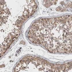
- Submitted by
- Atlas Antibodies (provider)
- Main image
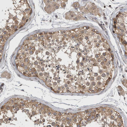
- Experimental details
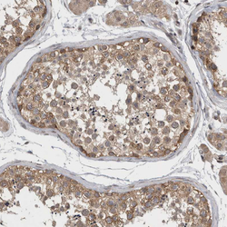
- Immunohistochemical staining of human testis shows moderate to strong cytoplasmic positivity in cells in seminiferous ducts.
- Sample type
- HUMAN
- Submitted by
- Atlas Antibodies (provider)
- Main image

- Experimental details
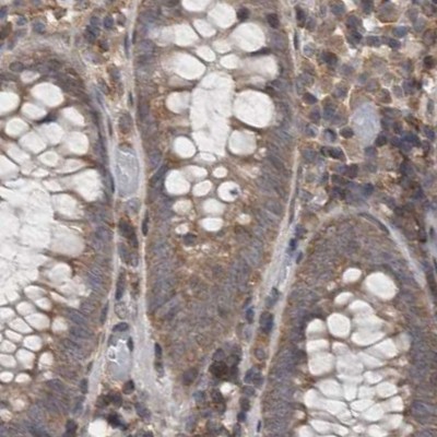
- Immunohistochemical staining of human colon using Anti-ATXN2 antibody HPA020339.
- Sample type
- HUMAN
- Submitted by
- Atlas Antibodies (provider)
- Main image
- Experimental details
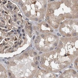
- Immunohistochemical staining of human kidney using Anti-ATXN2 antibody HPA020339.
- Sample type
- HUMAN

- Submitted by
- Atlas Antibodies (provider)
- Main image
- Experimental details
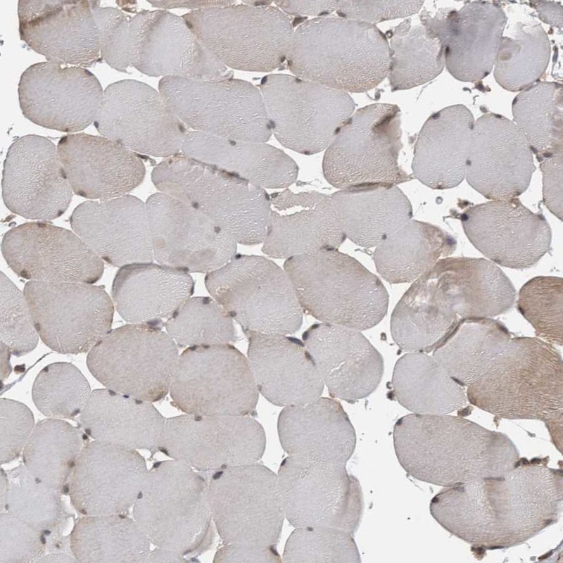
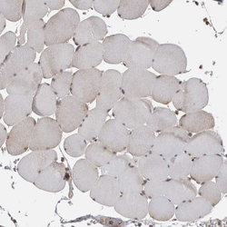
- Immunohistochemical staining of human skeletal muscle shows very weak cytoplasmic positivity in myocytes.
- Sample type
- HUMAN
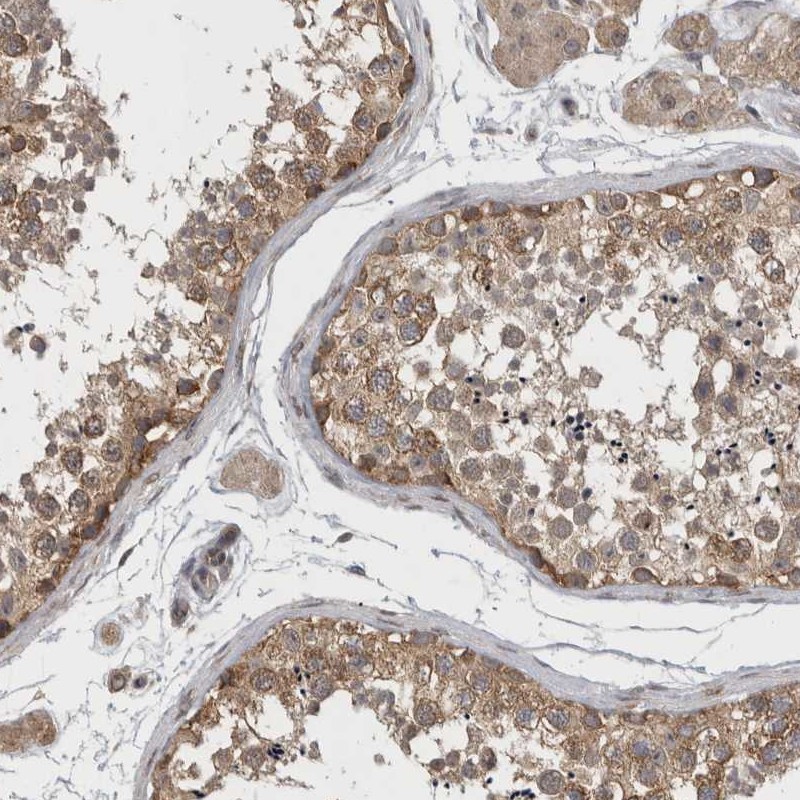
- Submitted by
- Atlas Antibodies (provider)
- Main image
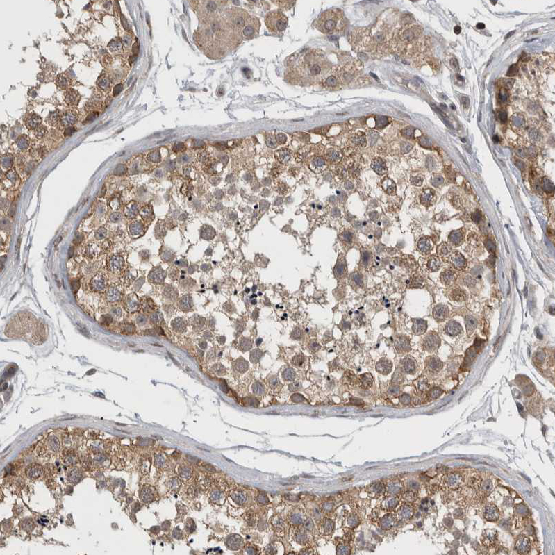
- Experimental details
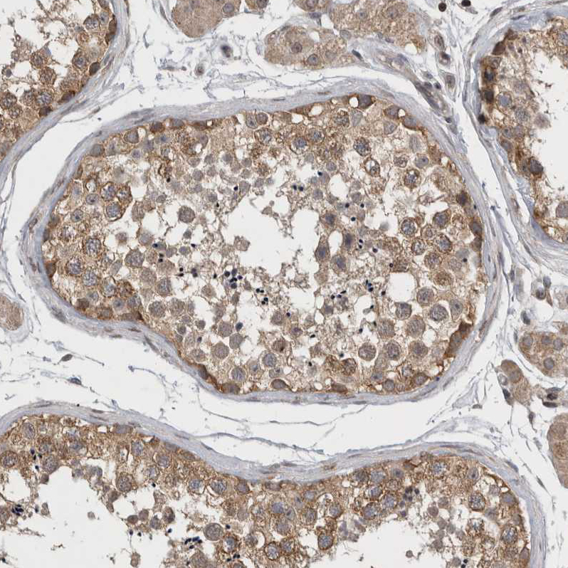
- Immunohistochemical staining of human testis shows moderate cytoplasmic positivity in cells in seminiferous ducts.
- Sample type
- HUMAN
- Submitted by
- Atlas Antibodies (provider)
- Main image
- Experimental details
- Immunohistochemical staining of human cerebellum shows strong cytoplasmic positivity in Purkinje cells.
- Sample type
- HUMAN

- Submitted by
- Atlas Antibodies (provider)
- Main image
- Experimental details
- Immunohistochemical staining of human cerebral cortex shows moderate to strong cytoplasmic positivity in neurons.
- Sample type
- HUMAN
