 Explore
 Validate
 Learn
Learn Western blot
Antibody data
- Antibody Data
- Antigen structure
- References [0]
- Comments [0]
- Validations
- Western blot [4]
- Immunocytochemistry [1]
- Immunohistochemistry [2]
Submit
Validation data
Reference
Comment
Report error
- Product number
- PA5-34638 - Provider product page

- Provider
- Invitrogen Antibodies
- Product name
- Urokinase Polyclonal Antibody
- Antibody type
- Polyclonal
- Antigen
- Recombinant protein fragment
- Description
- Recommended positive controls: H1299, HCT116, PC3 conditioned medium. Predicted reactivity: Dog (82%), Pig (82%), Rabbit (86%), Sheep (81%). IHC notes, Requires antigen retrieval using heat mediated 10mM Citrate buffer (pH6.0) or Tris-EDTA buffer (pH8.0) Store product as a concentrated solution. Centrifuge briefly prior to opening the vial.
- Reactivity
- Human
- Host
- Rabbit
- Isotype
- IgG
- Vial size
- 100 µL
- Concentration
- 0.4 mg/mL
- Storage
- Store at 4°C short term. For long term storage, store at -20°C, avoiding freeze/thaw cycles.
No comments: Submit comment
Supportive validation
- Submitted by
- Invitrogen Antibodies (provider)
- Main image

- Experimental details
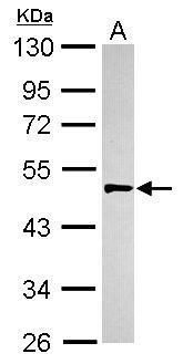
- Western blot analysis of Urokinase using 30 µg of HepG2 lysate. Samples were loaded onto a 10% SDS-PAGE gel and probed with a Urokinase polyclonal antibody (Product # PA5-34638) at a dilution of 1:500.
- Submitted by
- Invitrogen Antibodies (provider)
- Main image

- Experimental details
- PC-3 whole cell extract and conditioned medium (30 µg) were separated by 10% SDS-PAGE, and the membrane was blotted with Urokinase Polyclonal Antibody (Product # PA5-34638) diluted at 1:500.

- Submitted by
- Invitrogen Antibodies (provider)
- Main image

- Experimental details
- Western Blot analysis of Urokinase was performed by separating 30 µg of various whole cell extracts by 10% SDS-PAGE. Proteins were transferred to a membrane and probed with a Urokinase Polyclonal Antibody (Product # PA5-34638) at a dilution of 1:500.

- Submitted by
- Invitrogen Antibodies (provider)
- Main image

- Experimental details
- Western blot was performed using Anti-Urokinase Polyclonal Antibody (Product # PA5-34638) and a 50 k Da band corresponding to Urokinase was observed in PC-3 and DU 145 but not in LNCaP upon treatment with the protein secretory blockers, PTI and FLI-06. Whole cell extracts (30 µg lysate) of PC-3 (Lane 1), PC-3 treated with PTI (1X for 4 hours) (Lane 2), PC-3 treated with FLI-06 (10uM for 4 hours) (Lane 3), DU 145 (Lane 4), DU 145 treated with PTI (1X for 4 hours) (Lane 5), DU 145 treated with FLI-06 (10uM for 4 hours) (Lane 6), LNCaP (Lane 7), LNCaP treated with PTI (1X for 4 hours) (Lane 8) and LNCaP treated with FLI-06 (10uM for 4 hours) (Lane 9) were electrophoresed using NuPAGE™ 10% Bis-Tris Protein Gel (Product # NP0301BOX). Resolved proteins were then transferred onto a Nitrocellulose membrane (Product # IB23001) by iBlot® 2 Dry Blotting System (Product # IB21001). The blot was probed with the primary antibody (1:1000 dilution) and detected by chemiluminescence with Goat anti-Rabbit IgG (H+L) Superclonal™ Recombinant Secondary Antibody, HRP (Product # A27036, 1:4000 dilution) using the iBright FL 1000 (Product # A32752). ECL detection was performed using Novex® ECL Chemiluminescent Substrate Reagent Kit (Product # WP20005). Entrapment of Urokinase using PTI and FLI-06 increases intracellular accumulation in PC-3 and DU 145 but not in LNCaP which is reported to be negative.

Supportive validation
- Submitted by
- Invitrogen Antibodies (provider)
- Main image

- Experimental details
- Immunofluorescence analysis of Urokinase was performed using 70% confluent log phase PC-3 and LNCaP, control and PTI treated, cells. The cells were fixed with 4% paraformaldehyde for 10 minutes, permeabilized with 0.1% Triton™ X-100 for 15 minutes, and blocked with 2% BSA for 45 minutes at room temperature. The cells were labeled with Urokinase Polyclonal Antibody (Product # PA5-34638) at 1:100 dilution in 0.1% BSA, incubated at 4 degree celsius overnight and then labeled with Donkey anti-Rabbit IgG (H+L) Highly Cross-Adsorbed Secondary Antibody, Alexa Fluor Plus 488 (Product # A32790), (1:2000 dilution), for 45 minutes at room temperature (Panel a, e, i: Green). Nuclei (Panel b, f, j: Blue) were stained with ProLong™ Diamond Antifade Mountant with DAPI (Product # P36962). F-actin (Panel c, g, k: Blue) was stained with Rhodamine Phalloidin (Product # R415, 1:300). Panel d represents the merged image of untreated PC-3 cells showing faint staining for Urokinase that is enhanced upon PTI treatment (Panel h). Panel l represents LNCaP cells showing no staining for Urokinase upon PTI treatment. The images were captured at 60X magnification.

Supportive validation
- Submitted by
- Invitrogen Antibodies (provider)
- Main image
- Experimental details
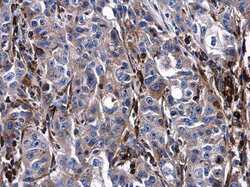
- Immunohistochemistry (Paraffin) analysis of Urokinase was performed in paraffin-embedded human esophageal carcinoma tissue using Urokinase Polyclonal Antibody (Product # PA5-34638) at a dilution of 1:500.

- Submitted by
- Invitrogen Antibodies (provider)
- Main image
- Experimental details
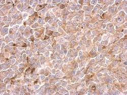
- Urokinase Polyclonal Antibody detects PLAU protein at cytosol on H1299 xenograft by immunohistochemical analysis. Sample: Paraffin-embedded H1299 xenograft. Urokinase Polyclonal Antibody (Product # PA5-34638) dilution: 1:500. Antigen Retrieval: EDTA based buffer, pH 8.0, 15 min.
