 Explore
 Validate
 Learn
Learn Western blot
 Flow cytometry
Antibody data
- Antibody Data
- Antigen structure
- References [1]
- Comments [0]
- Validations
- Western blot [1]
- Immunocytochemistry [1]
- Immunohistochemistry [1]
Submit
Validation data
Reference
Comment
Report error
- Product number
- GTX16827 - Provider product page

- Provider
- GeneTex
- Product name
- P2X7 antibody
- Antibody type
- Polyclonal
- Reactivity
- Human, Mouse, Rat
- Host
- Rabbit
Submitted references ATP/P2X7 receptor signaling as a potential anti-inflammatory target of natural polyphenols.
Nuka E, Ohnishi K, Terao J, Kawai Y
PloS one 2018;13(9):e0204229
PloS one 2018;13(9):e0204229
No comments: Submit comment
Supportive validation
- Submitted by
- GeneTex (provider)
- Main image

- Experimental details
- Anti-P2X7_Receptor_(extracellular) - Western blot analysis of rat brain membranes (lanes 1,5) and human cell lines; K562 (lanes 2 and 6); WEHI-231 (lanes 3 and 7) and HL-60 (lanes 4 and 8): 1-4. Anti-P2X7 Receptor (extracellular) antibody, (1:200). 5-8. Anti-P2X7 Receptor (extracellular) antibody, preincubated with the control peptide antigen.

Supportive validation
- Submitted by
- GeneTex (provider)
- Main image

- Experimental details
- Anti-P2X7_Receptor_(extracellular) - Expression of P2X7 in rat RBL cells Immunocytochemical staining of intact living rat basophilic leukemia (RBL) cells. A. Extracellular staining of cells using Anti-P2X7 Receptor (extracellular) antibody, (1:100) followed by goat anti-rabbit-AlexaFluor-594 secondary antibody (red). B. Live view of the cells.

Supportive validation
- Submitted by
- GeneTex (provider)
- Main image
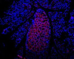
- Experimental details
- Anti-P2X7_Receptor_(extracellular) - Expression of P2X7 in rat pancreas Immunohistochemical staining of rat pancreas frozen section using Anti-P2X7 Receptor (extracellular) antibody, (1:100), followed by mouse anti-rabbit-AlexaFluor-594 secondary antibody (red). Staining is present in endocrine cells of the Isle of Langerhans. Hoechst 33342 is used as the counterstain.
