Explore
 Validate
 Learn
Learn Western blot
 Blocking/Neutralizing
Antibody data
- Antibody Data
- Antigen structure
- References [9]
- Comments [0]
- Validations
- Western blot [2]
- ELISA [1]
- Immunohistochemistry [1]
Submit
Validation data
Reference
Comment
Report error
- Product number
- AF156 - Provider product page

- Provider
- Novus Biologicals
- Product name
- Goat Polyclonal PD-L1 Antibody
- Antibody type
- Polyclonal
- Description
- Antigen Affinity-purified. Detects human PD-L1/B7-H1 in direct ELISAs and Western blots. In direct ELISAs, less than 15% cross-reactivity with recombinant mouse PD-L1/B7-H1 is observed and less than 1% cross-reactivity with recombinant human PD-L2 is observed.
- Reactivity
- Human
- Host
- Goat
- Conjugate
- Unconjugated
- Isotype
- IgG
- Vial size
- 100 ug
- Concentration
- LYOPH
- Storage
- Use a manual defrost freezer and avoid repeated freeze-thaw cycles. 12 months from date of receipt, -20 to -70 degreesC as supplied. 1 month, 2 to 8 degreesC under sterile conditions after reconstitution. 6 months, -20 to -70 degreesC under sterile conditions after reconstitution.
Submitted references Restoration of T Cell function in multi-drug resistant bacterial sepsis after interleukin-7, anti-PD-L1, and OX-40 administration.
Immunological Properties of Human Embryonic Stem Cell-Derived Retinal Pigment Epithelial Cells.
Hepatitis C Virus Induces MDSCs-Like Monocytes through TLR2/PI3K/AKT/STAT3 Signaling.
Mesenchymal Stromal Cell Secretion of Programmed Death-1 Ligands Regulates T Cell Mediated Immunosuppression.
Blocking the PD-1/PD-L1 pathway in glioma: a potential new treatment strategy.
Soluble co-signaling molecules predict long-term graft outcome in kidney-transplanted patients.
12-O-tetradecanoyl phorbol 13-acetate induces the expression of B7-DC, -H1, -H2, and -H3 in K562 cells.
Aberrant regulation of synovial T cell activation by soluble costimulatory molecules in rheumatoid arthritis.
Aberrant regulation of synovial T cell activation by soluble costimulatory molecules in rheumatoid arthritis.
Thampy LK, Remy KE, Walton AH, Hong Z, Liu K, Liu R, Yi V, Burnham CD, Hotchkiss RS
PloS one 2018;13(6):e0199497
PloS one 2018;13(6):e0199497
Immunological Properties of Human Embryonic Stem Cell-Derived Retinal Pigment Epithelial Cells.
Idelson M, Alper R, Obolensky A, Yachimovich-Cohen N, Rachmilewitz J, Ejzenberg A, Beider E, Banin E, Reubinoff B
Stem cell reports 2018 Sep 11;11(3):681-695
Stem cell reports 2018 Sep 11;11(3):681-695
Hepatitis C Virus Induces MDSCs-Like Monocytes through TLR2/PI3K/AKT/STAT3 Signaling.
Zhai N, Li H, Song H, Yang Y, Cui A, Li T, Niu J, Crispe IN, Su L, Tu Z
PloS one 2017;12(1):e0170516
PloS one 2017;12(1):e0170516
Mesenchymal Stromal Cell Secretion of Programmed Death-1 Ligands Regulates T Cell Mediated Immunosuppression.
Davies LC, Heldring N, Kadri N, Le Blanc K
Stem cells (Dayton, Ohio) 2017 Mar;35(3):766-776
Stem cells (Dayton, Ohio) 2017 Mar;35(3):766-776
Blocking the PD-1/PD-L1 pathway in glioma: a potential new treatment strategy.
Xue S, Hu M, Iyer V, Yu J
Journal of hematology & oncology 2017 Apr 7;10(1):81
Journal of hematology & oncology 2017 Apr 7;10(1):81
Soluble co-signaling molecules predict long-term graft outcome in kidney-transplanted patients.
Melendreras SG, Martínez-Camblor P, Menéndez A, Bravo-Mendoza C, González-Vidal A, Coto E, Díaz-Corte C, Ruiz-Ortega M, López-Larrea C, Suárez-Álvarez B
PloS one 2014;9(12):e113396
PloS one 2014;9(12):e113396
12-O-tetradecanoyl phorbol 13-acetate induces the expression of B7-DC, -H1, -H2, and -H3 in K562 cells.
Jang BC, Park YK, Choi IH, Kim SP, Hwang JB, Baek WK, Suh MH, Mun KC, Suh SI
International journal of oncology 2007 Dec;31(6):1439-47
International journal of oncology 2007 Dec;31(6):1439-47
Aberrant regulation of synovial T cell activation by soluble costimulatory molecules in rheumatoid arthritis.
Wan B, Nie H, Liu A, Feng G, He D, Xu R, Zhang Q, Dong C, Zhang JZ
Journal of immunology (Baltimore, Md. : 1950) 2006 Dec 15;177(12):8844-50
Journal of immunology (Baltimore, Md. : 1950) 2006 Dec 15;177(12):8844-50
No comments: Submit comment
Supportive validation
- Submitted by
- Novus Biologicals (provider)
- Main image

- Experimental details
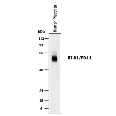
- Detection of Human PD-L1/B7-H1 by Western Blot. Western blot shows lysates of human placenta tissue. PVDF membrane was probed with 2 µg/mL of Goat Anti-Human PD-L1/B7-H1 Antigen Affinity-purified Polyclonal Antibody (Catalog # AF156) followed by HRP-conjugated Anti-Goat IgG Secondary Antibody (Catalog # HAF017). A specific band was detected for PD-L1/B7-H1 at approximately 50-55 kDa (as indicated). This experiment was conducted under reducing conditions and using Immunoblot Buffer Group 1.
- Submitted by
- Novus Biologicals (provider)
- Main image

- Experimental details
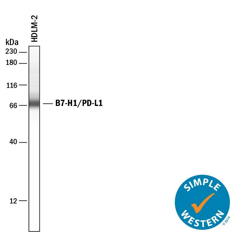
- Detection of Human PD-L1/B7-H1 by Simple WesternTM. Detection of Human PD-L1/B7-H1 by Simple WesternTM.
Supportive validation
- Submitted by
- Novus Biologicals (provider)
- Main image

- Experimental details
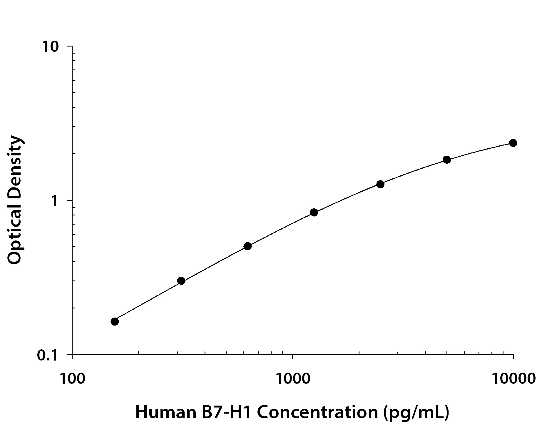
- Human PD-L1/B7-H1 ELISA Standard Curve. Human PD-L1/B7-H1 ELISA Standard Curve.
Supportive validation
- Submitted by
- Novus Biologicals (provider)
- Main image
- Experimental details
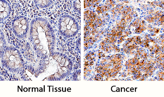
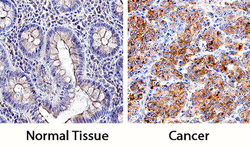
- PD-L1/B7-H1 in Human Colon and Colon Cancer Tissue. PD-L1/B7-H1 was detected in immersion fixed paraffin-embedded sections of normal human colon (left panel) and human colon cancer tissue (right panel) using Goat Anti-Human PD-L1/B7-H1 Antigen Affinity-purified Polyclonal Antibody (Catalog # AF156) at 5 µg/mL overnight at 4 °C. Tissue was stained using the Anti-Goat HRP-DAB Cell & Tissue Staining Kit (brown; Catalog # CTS008) and counterstained with hematoxylin (blue). Specific staining was localized to cell membranes and cytoplasm. View our protocol for Chromogenic IHC Staining of Paraffin-embedded Tissue Sections.