 Explore
 Validate
 Learn
Learn Western blot
Antibody data
- Antibody Data
- Antigen structure
- References [2]
- Comments [0]
- Validations
- Western blot [6]
- Immunohistochemistry [1]
- Other assay [2]
Submit
Validation data
Reference
Comment
Report error
- Product number
- PA1-46217 - Provider product page

- Provider
- Invitrogen Antibodies
- Product name
- STIM1 Polyclonal Antibody
- Antibody type
- Polyclonal
- Antigen
- Synthetic peptide
- Description
- The target sequence has 93% sequence homology with porcine and 79% sequence homology with xenopus. Suggested positive control: antigen standard for STIM1 (transient overexpression lysate), HeLa whole cell lysate.
- Reactivity
- Human, Mouse, Rat, Bovine, Canine
- Host
- Rabbit
- Isotype
- IgG
- Vial size
- 100 µL
- Concentration
- 1 mg/mL
- Storage
- Store at 4°C short term. For long term storage, store at -20°C, avoiding freeze/thaw cycles.
Submitted references X-ray irradiation triggers immune response in human T-lymphocytes via store-operated Ca2+ entry and NFAT activation.
Functional culture and in vitro genetic and small-molecule manipulation of adult mouse cardiomyocytes.
Tandl D, Sponagel T, Alansary D, Fuck S, Smit T, Hehlgans S, Jakob B, Fournier C, Niemeyer BA, Rödel F, Roth B, Moroni A, Thiel G
The Journal of general physiology 2022 May 2;154(5)
The Journal of general physiology 2022 May 2;154(5)
Functional culture and in vitro genetic and small-molecule manipulation of adult mouse cardiomyocytes.
Callaghan NI, Lee SH, Hadipour-Lakmehsari S, Lee XA, Ahsan Siraj M, Driouchi A, Yip CM, Husain M, Simmons CA, Gramolini AO
Communications biology 2020 May 11;3(1):229
Communications biology 2020 May 11;3(1):229
No comments: Submit comment
Supportive validation
- Submitted by
- Invitrogen Antibodies (provider)
- Main image
- Experimental details
- Western blot analysis of STIM1 in HeLa whole cell extracts using a polyclonal antibody (Product # PA1-46217).

- Submitted by
- Invitrogen Antibodies (provider)
- Main image

- Experimental details
- Western blot analysis of STIM1 using a polyclonal antibody (Product # PA1-46217).

- Submitted by
- Invitrogen Antibodies (provider)
- Main image
- Experimental details
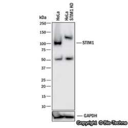
- Knockout validation by Western blot analysis of STIM1 in lysates of HeLa human cervical epithelial carcinoma parental cell line and Stim-1 knockout (KO) HeLa cell line. Samples were incubated in STIM1 polyclonal antibody (Product # PA1-46217) using a dilution of 1:1000 followed by a HRP-conjugated Anti-Rabbit IgG secondary antibody. Specific band was detected for Stim-1 at approximately 90 kDa (as indicated) in the parental HeLa cell line, but is not detectable in the knockout HeLa cell line. This experiment was conducted under reducing conditions.

- Submitted by
- Invitrogen Antibodies (provider)
- Main image

- Experimental details
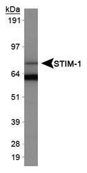
- Western blot analysis of STIM1 in HeLa whole cell extracts. Sample was incubated in STIM1 polyclonal antibody (Product # PA1-46217).

- Submitted by
- Invitrogen Antibodies (provider)
- Main image
- Experimental details
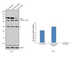
- Knockdown of STIM1 was achieved by transfecting PANC-1 with STIM1 specific siRNAs (Silencer® select Product # s531229, s531227). Western blot analysis (Fig. a) was performed using whole cell extracts from the STIM1 knockdown cells (Lane 3), non-specific scrambled siRNA transfected cells (Lane 2) and untransfected cells (Lane 1). The blot was probed with STIM1 Polyclonal Antibody (Product # PA1-46217, 1 µg/ml) and Goat anti-Rabbit IgG (H+L), Superclonal™ Recombinant Secondary Antibody, HRP (Product # A27036, 0.25µg/ml, 1:4000 dilution). Densitometric analysis of this western blot is shown in histogram (Fig. b). Decrease in signal (83 kDa isoform) upon siRNA mediated knock down confirms that antibody is specific to STIM1. (Note: An uncharacterized band was observed at 50 kDa).

- Submitted by
- Invitrogen Antibodies (provider)
- Main image
- Experimental details
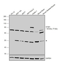
- Western blot was performed using Anti-STIM1 Polyclonal Antibody (Product # PA1-46217) and bands corresponding to 83kDa and 77kDa of STIM1 were observed across all the cell lines and tissues tested along with an uncharacterized band (*) at ~50kDa across the panel. Whole cell extracts (30 µg lysate) of PC-12 (Lane 1), MCF 10A (Lane 2), MCF7 (Lane 3), MIA PaCa-2 (Lane 4), PANC-1 (Lane 5), tissue extracts (30ug lysate) of Mouse Brain (Lane 6) ad Mouse Skeletal Muscle (Lane 7) were electrophoresed using NuPAGE™ 10% Bis-Tris Protein Gel (Product # NP0302BOX). Resolved proteins were then transferred onto a nitrocellulose membrane (Product # IB23001) by iBlot® 2 Dry Blotting System (Product # IB21001).The blot was probed with the primary antibody (1 µg/ml) and detected by chemiluminescence with Goat anti-Rabbit IgG (H+L), Superclonal™ Recombinant Secondary Antibody, HRP (Product # A27036, 1:4000 dilution) using the iBright FL 1000 (Product # A32752). Chemiluminescent detection was performed using Novex® ECL Chemiluminescent Substrate Reagent Kit (Product # WP20005).

Supportive validation
- Submitted by
- Invitrogen Antibodies (provider)
- Main image

- Experimental details
- STIM-1 staining in Hela cells detected with a Dylight 488 labeled secondary antibody.

Supportive validation
- Submitted by
- Invitrogen Antibodies (provider)
- Main image

- Experimental details
- Fig. 1 Isolated adult murine cardiomyocytes (aCMs) cultured on Geltrex and treated with blebbistatin show sustained viability and retention of functional protein expression patterns up to 7 days post-isolation. a Confocal analysis of aCMs of alpha-actinin and RyR2, DHPRalpha, SERCA2a, and STIM1 (green) up to one week in culture. Nuclei were visualized with Hoechst 33342 (blue). Scale bars equal 20 um. b Immunoblots for Akt, pAkt, and alpha-tubulin of aCM lysates after 24 h culture on Geltrex-coated surfaces ( N = 3, p = 0.023). c Cell counting revealed the survival of viable rod-shaped (red) relative to rounded aCMs (blue) ( N = 3). All data expressed as mean +- SEM; N denotes biological replicates; significance indicated by *( p < 0.05).

- Submitted by
- Invitrogen Antibodies (provider)
- Main image

- Experimental details
- Figure S2. In resting Jurkat cells, STIM1 and Orai1 are located in the ER and PM, respectively. (A and C) Confocal images of cellular distribution of endogenous STIM1 and Orai1 in Jurkat cells (A) and PBMCs (C). PM and ER of cells (first row) were stained with CellMaskOrange and ER-tracker red, respectively. Images shown as false color in blue. The second row shows immunostaining of STIM1 (green) and Orai1 (magenta) and secondary antibody tagged with Alx488 and Alx647, respectively. An overlay of both channels is shown in right column. (B and D) Line plots for each marker were taken in positions report in merged images. Fluorescence intensity of either Orai1/PM (B) or STIM1/ER (D) were normalized to the highest value of each signal; the colors of line plots correspond to those in images. All scale bars, 10 mum. The antibodies for detecting STIM1 and Orai1 are specific. (E) Jurkat cells in which Orai1 was knocked out ( Fig. S2 ) generate no more signal in immunostaining with Orai1 antibody, while still producing signal with STIM1 antibody. (F) Staining of WT Jurkat cells in which either the primary STIM1 or Orai1 antibodies (top row) or the respective secondary antibodies (bottom row) were left out generated no appreciable fluorescent signals.
