 Explore
 Validate
 Learn
Learn Western blot
 Immunocytochemistry
Antibody data
- Antibody Data
- Antigen structure
- References [0]
- Comments [0]
- Validations
- Western blot [1]
- Immunohistochemistry [1]
Submit
Validation data
Reference
Comment
Report error
- Product number
- AP09082SU-N - Provider product page

- Provider
- Acris Antibodies GmbH
- Proper citation
- Acris Antibodies GmbH Cat#AP09082SU-N, RRID:AB_2035838
- Product name
- anti EIF2AK3 (601-1115)
- Antibody type
- Polyclonal
- Antigen
- Recombinant fusion protein from amino acids 601-1115 of Mouse deltaN PERK
- Reactivity
- Mouse
- Host
- Rabbit
- Isotype
- IgG
- Vial size
- 0.1 ml
- Concentration
- 85 mg/ml (by Refractometry)
No comments: Submit comment
Supportive validation
- Submitted by
- Acris Antibodies GmbH (provider)
- Main image
- Experimental details
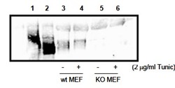
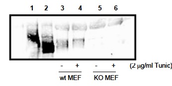
- Western blot analysis: AP09082SU-N PERK antibody staining of cell lysates. 300µg PERK over-expressing 293T cell lysate (Lanes 1 and 2), or 800µg wild type (Lanes 3 and 4), and PERK knock out (Lanes 5 and 6) MEF cell lysate were immunoprecipated with 15µl anti-PERK, followed by immunobloting with 1/1000 dilution of anti-PERK in 5% milk/TBST buffer. Lane 1, 293T cells over-expressing Myc-PERK wt, Lane 2 , 293T cells over-expressing Myc-PERK K618A. Personal Communication. A, Diehl, Univ. of Pennsylvania, Philadelphia, PA.
Supportive validation
- Submitted by
- Acris Antibodies GmbH (provider)
- Main image

- Experimental details
- Immunohistochemistry staining of Mouse mammary gland samples from lactating mice (L10) using AP09082SU-N PERK antibody. Positive staining signal observed in wild type Mouse sample with anti-PERK staining only (middle image), but not in the knock out mouse sample (Right image) and pre-immune serum staining (Left image). The anti-PERK was diluted 1/1,000 in 5% goat serum in PBS and allowed to incubate for 2h at RT in a humidified chamber. Personal Communication. A, Diehl, Univ. of Pennsylvania, Philadelphia, PA.
