 Explore
 Validate
 Learn
Learn Western blot
Antibody data
- Antibody Data
- Antigen structure
- References [0]
- Comments [0]
- Validations
- Western blot [1]
- Immunohistochemistry [2]
Submit
Validation data
Reference
Comment
Report error
- Product number
- TA328608 - Provider product page

- Provider
- OriGene
- Product name
- Rabbit Polyclonal Anti-M1 Muscarinic Receptor
- Antibody type
- Polyclonal
- Description
- Rabbit Polyclonal Anti-M1 Muscarinic Receptor
- Host
- Rabbit
- Conjugate
- Unconjugated
- Epitope
- CHRM1
- Antibody clone number
- NULL
- Vial size
- 200 µl
- Concentration
- NULL
No comments: Submit comment
Supportive validation
- Submitted by
- OriGene (provider)
- Main image
- Experimental details
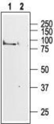
- Western blot analysis of rat brain membranes: 1. Anti-M1 Muscarinic Receptor antibody, (1:200). 2. Anti-M1 Muscarinic Receptor antibody, preincubated with the control fusion protein antigen.
- Validation comment
- WB

Supportive validation
- Submitted by
- OriGene (provider)
- Main image

- Experimental details
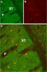
- Expression of M1 in rat striatum. Immunohistochemical staining of rat striatum (ST) using Anti-M1 Muscarinic Receptor antibody. A. M1 Muscarinic Receptor appears in the striatum (green). B. Staining of interneurons with mouse anti-parvalbumin (PV, red). C. Confocal merge of M1 Muscarinic Receptor and PV demonstrates localization of PV expressing neurons in the striatal matrix; not in striatal patches (P).
- Validation comment
- IHC

- Submitted by
- OriGene (provider)
- Main image
- Experimental details
- Expression of M1 in mouse striatum. Immunohistochemical staining of mouse striatum (ST) using Anti-M1 Muscarinic Receptor antibody. A. M1 Muscarinic Receptor appears in the striatum (green). B. Staining of interneurons with mouse anti-parvalbumin (PV, red). C. Confocal merge of M1 Muscarinic Receptor and PV demonstrates localization of PV expressing neurons in the striatal matrix; not in striatal patches (P).
- Validation comment
- IHC
