 Explore
 Validate
 Learn
LearnAF1987
antibody from R&D Systems
Targeting: CD59
16.3A5, EJ16, EJ30, EL32, G344, MIC11, MIN1, MIN2, MIN3, MSK21, p18-20
 Western blot
Antibody data
- Antibody Data
- Antigen structure
- References [2]
- Comments [0]
- Validations
- Western blot [2]
- Immunohistochemistry [1]
Submit
Validation data
Reference
Comment
Report error
- Product number
- AF1987 - Provider product page

- Provider
- R&D Systems
- Product name
- Human CD59 Antibody
- Antibody type
- Polyclonal
- Description
- Antigen Affinity-purified. Detects human CD59 in direct ELISAs and Western blots.
- Reactivity
- Human
- Host
- Goat
- Conjugate
- Unconjugated
- Antigen sequence
P13987- Isotype
- IgG
- Vial size
- 100 ug
- Concentration
- LYOPH
- Storage
- Use a manual defrost freezer and avoid repeated freeze-thaw cycles. 12 months from date of receipt, -20 to -70 °C as supplied. 1 month, 2 to 8 °C under sterile conditions after reconstitution. 6 months, -20 to -70 °C under sterile conditions after reconstitution.
Submitted references Apolipoprotein J/clusterin in human erythrocytes is involved in the molecular process of defected material disposal during vesiculation.
Angiotensin II controls occludin function and is required for blood brain barrier maintenance: relevance to multiple sclerosis.
Antonelou MH, Kriebardis AG, Stamoulis KE, Trougakos IP, Papassideri IS
PloS one 2011;6(10):e26033
PloS one 2011;6(10):e26033
Angiotensin II controls occludin function and is required for blood brain barrier maintenance: relevance to multiple sclerosis.
Wosik K, Cayrol R, Dodelet-Devillers A, Berthelet F, Bernard M, Moumdjian R, Bouthillier A, Reudelhuber TL, Prat A
The Journal of neuroscience : the official journal of the Society for Neuroscience 2007 Aug 22;27(34):9032-42
The Journal of neuroscience : the official journal of the Society for Neuroscience 2007 Aug 22;27(34):9032-42
No comments: Submit comment
Supportive validation
- Submitted by
- R&D Systems (provider)
- Main image

- Experimental details
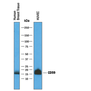
- Detection of Human CD59 by Western Blot. Western blot shows lysates of human breast tissue and HUVEC human umbilical vein endothelial cells. PVDF membrane was probed with 1 µg/mL of Goat Anti-Human CD59 Antigen Affinity-purified Polyclonal Antibody (Catalog # AF1987) followed by HRP-conjugated Anti-Goat IgG Secondary Antibody (Catalog # HAF019). A specific band was detected for CD59 at approximately 16 kDa (as indicated). This experiment was conducted under reducing conditions and using Immunoblot Buffer Group 1.
- Submitted by
- R&D Systems (provider)
- Main image
- Experimental details
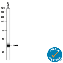
- Detection of Human CD59 by Simple WesternTM. Simple Western lane view shows lysates of HUVEC human umbilical vein endothelial cells, loaded at 0.2 mg/mL. A specific band was detected for CD59 at approximately 26 kDa (as indicated) using 50 µg/mL of Goat Anti-Human CD59 Antigen Affinity-purified Polyclonal Antibody (Catalog # AF1987) followed by 1:50 dilution of HRP-conjugated Anti-Goat IgG Secondary Antibody (Catalog # HAF109). This experiment was conducted under reducing conditions and using the 12-230 kDa separation system.

Supportive validation
- Submitted by
- R&D Systems (provider)
- Main image
- Experimental details
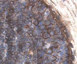
- CD59 in Human Breast. CD59 was detected in immersion fixed paraffin-embedded sections of human breast using 1.7 µg/mL Goat Anti-Human CD59 Antigen Affinity-purified Polyclonal Antibody (Catalog # AF1987) overnight at 4 °C. Tissue was stained with the Anti-Goat HRP-DAB Cell & Tissue Staining Kit (brown; Catalog # CTS008) and counterstained with hematoxylin (blue). View our protocol for Chromogenic IHC Staining of Paraffin-embedded Tissue Sections.
