Explore
 Validate
 Learn
Learn Western blot
 ELISA
ELISAAntibody data
- Antibody Data
- Antigen structure
- References [0]
- Comments [0]
- Validations
- Western blot [4]
- Immunocytochemistry [1]
- Immunohistochemistry [2]
- Chromatin Immunoprecipitation [1]
Submit
Validation data
Reference
Comment
Report error
- Product number
- MA5-17143 - Provider product page

- Provider
- Invitrogen Antibodies
- Product name
- PAX5 Monoclonal Antibody (7D3)
- Antibody type
- Monoclonal
- Antigen
- Purifed from natural sources
- Description
- MA5-17143 targets PAX5 in indirect ELISA, IHC and WB applications and shows reactivity with Human samples. The MA5-17143 immunogen is purified recombinant fragment of human PAX5 (amino acids: 235-382) expressed in E. Coli. MA5-17143 detects PAX5 which has a predicted molecular weight of approximately 45kDa.
- Reactivity
- Human
- Host
- Mouse
- Isotype
- IgG
- Antibody clone number
- 7D3
- Vial size
- 100 µg
- Concentration
- 1 mg/mL
- Storage
- Store at 4°C short term. For long term storage, store at -20°C, avoiding freeze/thaw cycles.
No comments: Submit comment
Supportive validation
- Submitted by
- Invitrogen Antibodies (provider)
- Main image

- Experimental details
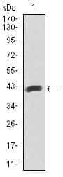
- Western blot analysis of PAX5 using a PAX5 monoclonal antibody (Product # MA5-17143) against a human PAX5 recombinant protein.

- Submitted by
- Invitrogen Antibodies (provider)
- Main image
- Experimental details
- Western blot analysis of PAX5 using a PAX5 monoclonal antibody (Product # MA5-17143) against a human PAX5 recombinant protein.

- Submitted by
- Invitrogen Antibodies (provider)
- Main image

- Experimental details
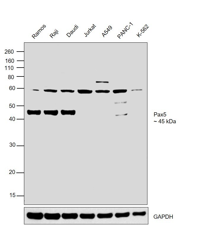
- Western blot was performed using Anti-PAX5 Monoclonal Antibody (7D3) (Product # MA5-17143) and a 45 kDa band corresponding to PAX5 was observed in Ramos, Raji and Daudi and not in Jurkat, A549, PANC-1 and K-562 along with an uncharacterized band at ~60 kDa. Modified whole cell extracts (1% SDS) (30 µg lysate) of Ramos (Lane 1), Raji (Lane 2), Daudi (Lane 3), Jurkat (Lane 4), A549 (Lane 5), PANC-1 (Lane 6) and K-562 (Lane 7) were electrophoresed using Novex® NuPAGE® 4-12% Bis-Tris Protein Gel (Product # NP0322BOX). Resolved proteins were then transferred onto a nitrocellulose membrane (Product # IB23001) by iBlot® 2 Dry Blotting System (Product # IB21001). The blot was probed with the primary antibody (1:1000 dilution) and detected by chemiluminescence with Goat anti-Mouse IgG (H+L), Superclonal™ Recombinant Secondary Antibody, HRP (Product # A28177, 1:4000 dilution) using the iBright FL 1000 (Product # A32752). Chemiluminescent detection was performed using Novex® ECL Chemiluminescent Substrate Reagent Kit (Product # WP20005).
- Submitted by
- Invitrogen Antibodies (provider)
- Main image

- Experimental details
- Western blot analysis of PAX5 using a PAX5 monoclonal antibody (Product # MA5-17143) against a human PAX5 recombinant protein.

Supportive validation
- Submitted by
- Invitrogen Antibodies (provider)
- Main image
- Experimental details
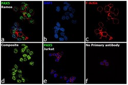
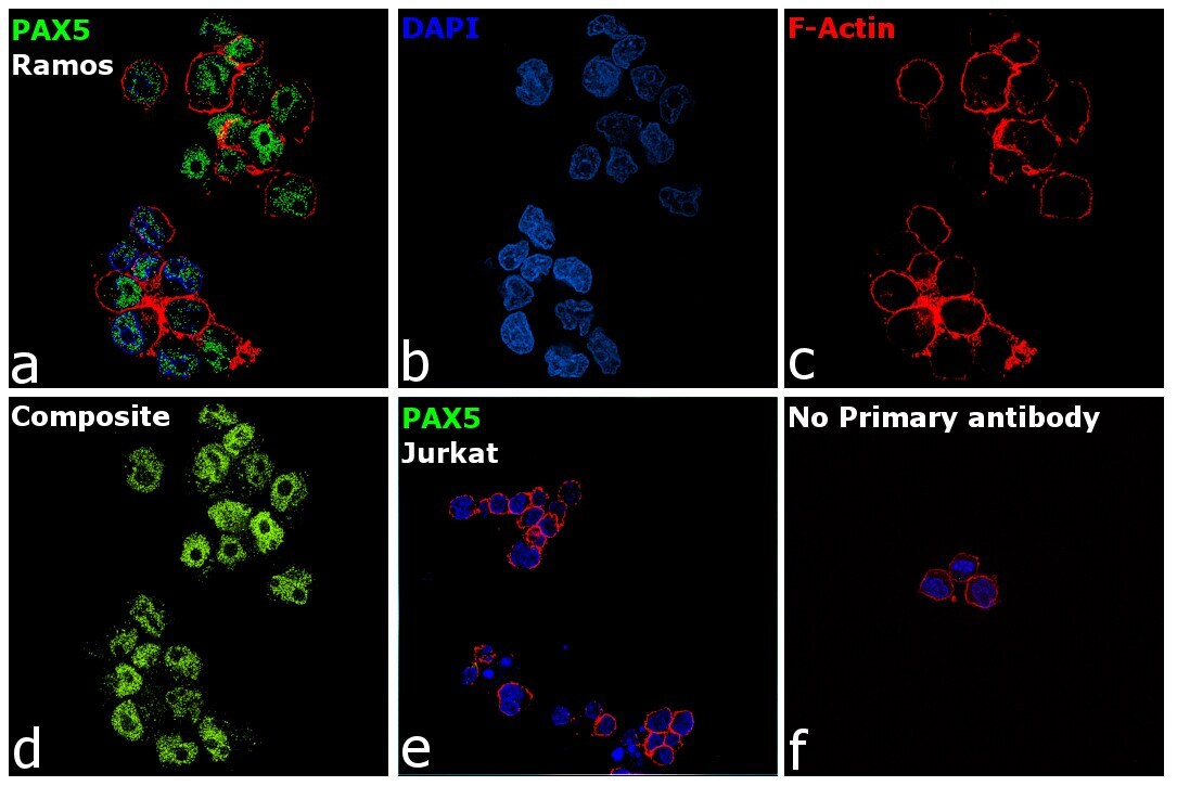
- Immunofluorescence analysis of PAX5 was performed using 70% confluent log phase Ramos cells. The cells were fixed with 4% Paraformaldehyde for 10 minutes, permeabilized with 0.1% Triton™ X-100 for 10 minutes, and blocked with 2% BSA for 10 minutes at room temperature. The cells were labeled with PAX5 Monoclonal Antibody (7D3) (Product # MA5-17143) at 1 µg/mL in 0.1% BSA, incubated at 4 degree Celsius overnight and then labeled with Goat anti-Mouse IgG (H+L), Superclonal™ Recombinant Secondary Antibody, Alexa Fluor 488 (Product # A28175) at a dilution of 1:2000 for 45 minutes at room temperature (Panel a: Green). Nuclei (Panel b: Blue) were stained with SlowFade® Gold Antifade Mountant with DAPI (Product # S36938). F-actin (Panel c: Red) was stained with Rhodamine Phalloidin (Product # R415, 1:300). Panel d represents the merged image showing nuclear localization. Panel e represents Jurkat cells having no expression of PAX5. Panel f represents control cells with no primary antibody to assess background. The images were captured at 60X magnification.
Supportive validation
- Submitted by
- Invitrogen Antibodies (provider)
- Main image
- Experimental details
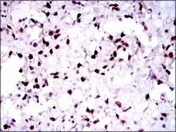
- Immunohistochemical analysis of paraffin-embedded brain tumors tissues using PAX5 monoclonal antibody (Product # MA5-17143) followed with DAB staining.

- Submitted by
- Invitrogen Antibodies (provider)
- Main image
- Experimental details
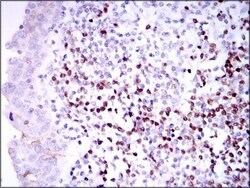
- Immunohistochemical analysis of paraffin-embedded endometrial cancer tissues using PAX5 monoclonal antibody (Product # MA5-17143) followed with DAB staining.

Supportive validation
- Submitted by
- Invitrogen Antibodies (provider)
- Main image
- Experimental details
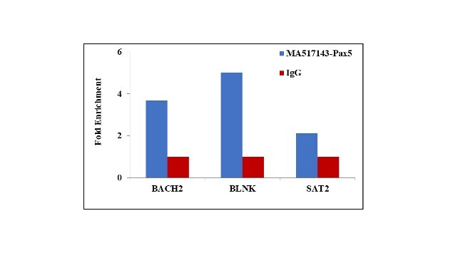
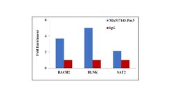
- Chromatin Immunoprecipitation (ChIP) assay of endogenous PAX5 protein using Anti-PAX5 Antibody: ChIP was performed using Anti-PAX5 Monoclonal Antibody (Product # MA5-17143, 5 µg) on sheared chromatin from Raji cells using the MAGnify ChIP System kit (Product # 49-2024). Normal Mouse IgG was used as a negative IP control. The purified DNA was analyzed by qPCR using primers binding to BACH2 and BLNK promoter and SAT2 satellite repeats. Data is presented as fold enrichment of the antibody signal versus the negative control IgG using the comparative CT method.