 Explore
 Validate
 Learn
Learn Western blot
 Flow cytometry
Antibody data
- Antibody Data
- Antigen structure
- References [2]
- Comments [0]
- Validations
- Western blot [2]
- Immunohistochemistry [1]
- Blocking/Neutralizing [1]
Submit
Validation data
Reference
Comment
Report error
- Product number
- AF382 - Provider product page

- Provider
- R&D Systems
- Product name
- Human CD27/TNFRSF7 Antibody
- Antibody type
- Polyclonal
- Description
- Antigen Affinity-purified. Detects human CD27 in direct ELISAs and Western blots. In direct ELISAs, approximately 25% cross-reactivity with recombinant mouse CD27 is observed and less than 1% cross-reactivity with recombinant human (rh) CD30, rh4-1BB, rhCD40, rhDR3, rhDR6, rhFas, rhGITR, rhHVEM, rhRANK, rhNGF R, rhsTNF RI, and rhsTNF RII is observed.
- Reactivity
- Human
- Host
- Goat
- Conjugate
- Unconjugated
- Antigen sequence
P26842- Isotype
- IgG
- Vial size
- 100 ug
- Concentration
- LYOPH
- Storage
- Use a manual defrost freezer and avoid repeated freeze-thaw cycles. 12 months from date of receipt, -20 to -70 °C as supplied. 1 month, 2 to 8 °C under sterile conditions after reconstitution. 6 months, -20 to -70 °C under sterile conditions after reconstitution.
Submitted references Blockade of protease-activated receptors on T cells correlates with altered proteolysis of CD27 by gingipains of Porphyromonas gingivalis.
IL-7 decreases IL-7 receptor alpha (CD127) expression and induces the shedding of CD127 by human CD8+ T cells.
Yun LW, Decarlo AA, Hunter N
Clinical and experimental immunology 2007 Nov;150(2):217-29
Clinical and experimental immunology 2007 Nov;150(2):217-29
IL-7 decreases IL-7 receptor alpha (CD127) expression and induces the shedding of CD127 by human CD8+ T cells.
Vranjkovic A, Crawley AM, Gee K, Kumar A, Angel JB
International immunology 2007 Dec;19(12):1329-39
International immunology 2007 Dec;19(12):1329-39
No comments: Submit comment
Supportive validation
- Submitted by
- R&D Systems (provider)
- Main image

- Experimental details
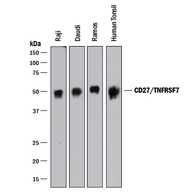
- Detection of Human CD27/TNFRSF7 by Western Blot. Western blot shows lysates of Raji human Burkitt's lymphoma cell line, Daudi human Burkitt's lymphoma cell line, Ramos human Burkitt's lymphoma cell line, and human tonsil tissue. PVDF membrane was probed with 1 µg/mL of Goat Anti-Human CD27/TNFRSF7 Antigen Affinity-purified Polyclonal Antibody (Catalog # AF382) followed by HRP-conjugated Anti-Goat IgG Secondary Antibody (Catalog # HAF017). A specific band was detected for CD27/ TNFRSF7 at approximately 50 kDa (as indicated). This experiment was conducted under reducing conditions and using Immunoblot Buffer Group 1.
- Submitted by
- R&D Systems (provider)
- Main image
- Experimental details
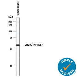
- Detection of Human CD27/TNFRSF7 by Simple WesternTM. Simple Western lane view shows lysates of human tonsil tissue, loaded at 0.2 mg/mL. A specific band was detected for CD27/TNFRSF7 at approximately 49 kDa (as indicated) using 20 µg/mL of Goat Anti-Human CD27/ TNFRSF7 Antigen Affinity-purified Polyclonal Antibody (Catalog # AF382) followed by 1:50 dilution of HRP-conjugated Anti-Goat IgG Secondary Antibody (Catalog # HAF109). This experiment was conducted under reducing conditions and using the 12-230 kDa separation system.

Supportive validation
- Submitted by
- R&D Systems (provider)
- Main image
- Experimental details
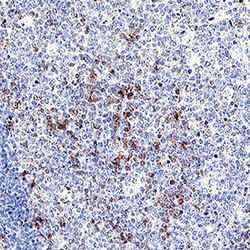
- CD27/TNFRSF7 in Human Tonsil. CD27/TNFRSF7 was detected in immersion fixed paraffin-embedded sections of human tonsil using Goat Anti-Human CD27/TNFRSF7 Antigen Affinity-purified Polyclonal Antibody (Catalog # AF382) at 3 µg/mL overnight at 4 °C. Tissue was stained using the Anti-Goat HRP-DAB Cell & Tissue Staining Kit (brown; Catalog # CTS008) and counterstained with hematoxylin (blue). Specific staining was localized to lymphocytes in germinal center. View our protocol for Chromogenic IHC Staining of Paraffin-embedded Tissue Sections.

Supportive validation
- Submitted by
- R&D Systems (provider)
- Main image
- Experimental details
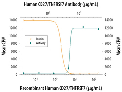
- CD27/TNFRSF7 Inhibition of Cell Proliferation and Neutralization by Human CD27/TNFRSF7 Antibody. In the presence of sub-optimal amounts of Mouse CD3 epsilon Monoclonal Anti-body (Catalog # MAB484) and Recombinant Mouse CD27 Ligand (10 µg/mL, Catalog # 783-CL), Recom-binant Human CD27 Fc Chimera (Catalog # 382-CD) inhibits proliferation in mouse splenic T cells in a dose-dependent manner (orange line). Under these conditions, inhibition of proliferation elicited by Recombinant Human CD27 Fc Chimera (3 µg/mL) is neutralized (green line) by increasing concentrations of Goat Anti-Human CD27 Antigen Affinity-purified Polyclonal Antibody (Catalog # AF382). The ND50 is typically 2.25-9.0 µg/mL.
