 Explore
 Validate
 Learn
Learn Western blot
 Immunocytochemistry
Antibody data
- Antibody Data
- Antigen structure
- References [2]
- Comments [0]
- Validations
- Immunocytochemistry [1]
Submit
Validation data
Reference
Comment
Report error
- Product number
- AF440 - Provider product page

- Provider
- R&D Systems
- Product name
- Mouse CD40/TNFRSF5 Antibody
- Antibody type
- Polyclonal
- Description
- Immunogen affinity purified. Detects mouse CD40/TNFRSF5 in direct ELISAs and Western blots. In Western blots, less than 1% cross-reactivity with recombinant human CD40 is observed. For B cell activation, the use of R&D Systems monoclonal anti-mouse CD40 antibody (Catalog # MAB440) is recommended.
- Reactivity
- Mouse
- Host
- Goat
- Conjugate
- Unconjugated
- Antigen sequence
P27512- Isotype
- IgG
- Vial size
- 100 ug
- Concentration
- LYOPH
- Storage
- Use a manual defrost freezer and avoid repeated freeze-thaw cycles. 12 months from date of receipt, -20 to -70 °C as supplied. 1 month, 2 to 8 °C under sterile conditions after reconstitution. 6 months, -20 to -70 °C under sterile conditions after reconstitution.
Submitted references Permissiveness to form pluripotent stem cells may be an evolutionarily derived characteristic in Mus musculus.
A PPARγ-dependent miR-424/503-CD40 axis regulates inflammation mediated angiogenesis.
Garbutt TA, Konneker TI, Konganti K, Hillhouse AE, Swift-Haire F, Jones A, Phelps D, Aylor DL, Threadgill DW
Scientific reports 2018 Oct 2;8(1):14706
Scientific reports 2018 Oct 2;8(1):14706
A PPARγ-dependent miR-424/503-CD40 axis regulates inflammation mediated angiogenesis.
Lee A, Papangeli I, Park Y, Jeong HN, Choi J, Kang H, Jo HN, Kim J, Chun HJ
Scientific reports 2017 May 31;7(1):2528
Scientific reports 2017 May 31;7(1):2528
No comments: Submit comment
Supportive validation
- Submitted by
- R&D Systems (provider)
- Main image
- Experimental details
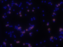
- CD40/TNFRSF5 in Mouse Splenocytes. CD40/TNFRSF5 was detected in immersion fixed mouse splenocytes using 10 µg/mL Goat Anti-Mouse CD40/TNFRSF5 Antigen Affinity-purified Polyclonal Antibody (Catalog # AF440) for 3 hours at room temperature. Cells were stained with the NorthernLights™ 557-conjugated Anti-Goat IgG Secondary Antibody (red; Catalog # NL001) and counterstained with DAPI (blue). View our protocol for Fluorescent ICC Staining of Non-adherent Cells.
