 Explore
 Validate
 Learn
Learn Western blot
 Immunohistochemistry
Antibody data
- Antibody Data
- Antigen structure
- References [1]
- Comments [0]
- Validations
- Western blot [1]
- Immunocytochemistry [2]
- Immunohistochemistry [5]
Submit
Validation data
Reference
Comment
Report error
- Product number
- HPA024120 - Provider product page

- Provider
- Atlas Antibodies
- Proper citation
- Atlas Antibodies Cat#HPA024120, RRID:AB_1858191
- Product name
- Anti-TOP2B
- Antibody type
- Polyclonal
- Reactivity
- Human, Mouse, Rat
- Host
- Rabbit
- Conjugate
- Unconjugated
- Antigen sequence
LWKEDLAAFVEELDKVESQEREDVLAGMSGKAIKG
KVGKPKVKKLQLEETMPSPYGRRIIPEITAMKADA
SKKLLKKKKGDLDTAAVKVEFDEEFSGAPVEGAGE
EALTPSVPINKGPKPKREKKEPGTRVRKTPTSSGK
PSA- Isotype
- IgG
- Vial size
- 100 µl
- Storage
- Store at +4°C for short term storage. Long time storage is recommended at -20°C.
Submitted references Uncovering Hidden Layers of Cell Cycle Regulation through Integrative Multi-omic Analysis.
Aviner R, Shenoy A, Elroy-Stein O, Geiger T
PLoS genetics 2015 Oct;11(10):e1005554
PLoS genetics 2015 Oct;11(10):e1005554
No comments: Submit comment
Enhanced validation
- Submitted by
- Atlas Antibodies (provider)
- Enhanced method
- Genetic validation
- Main image
- Experimental details
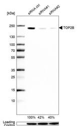
- Western blot analysis in U2OS cells transfected with control siRNA, target specific siRNA probe #1 and #2, using Anti-TOP2B antibody. Remaining relative intensity is presented. Loading control: Anti-GAPDH.

Enhanced validation
Supportive validation
- Submitted by
- 55af80e3e0991
- Enhanced method
- Genetic validation
- Main image
- Experimental details
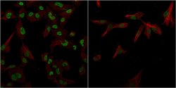
- Confocal images of immunofluorescently stained human U-2 OS cells.The protein TOP2B is shown in green and the microtubules in red. The image to the left show cells transfected with control siRNA and the image to the right show cells where TOP2B has been downregulated with specific siRNA.
- Sample type
- U-2 OS cells
- Primary Ab dilution
- 1:105
- Secondary Ab
- Secondary Ab
- Secondary Ab dilution
- 1:800
- Knockdown/Genetic Approaches Application
- Immunocytochemistry
Supportive validation
- Submitted by
- Atlas Antibodies (provider)
- Main image

- Experimental details
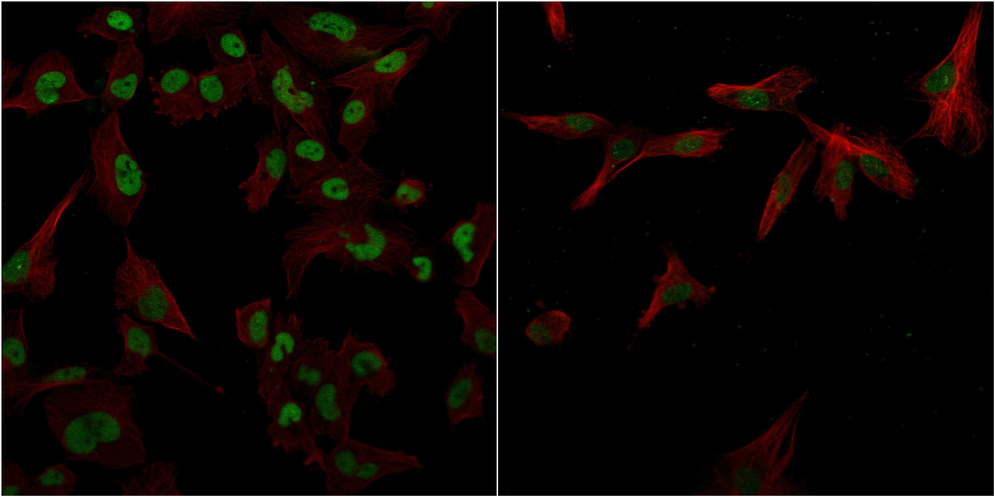
- Immunofluorescent staining of human cell line U-251 MG shows localization to nucleoplasm.
- Sample type
- HUMAN

Supportive validation
- Submitted by
- Atlas Antibodies (provider)
- Main image

- Experimental details
- Immunohistochemical staining of human pancreas shows moderate nuclear positivity.

- Submitted by
- Atlas Antibodies (provider)
- Main image
- Experimental details
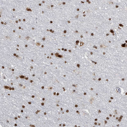
- Immunohistochemical staining of human cerebral cortex shows strong nuclear positivity in neurons.
- Sample type
- HUMAN

- Submitted by
- Atlas Antibodies (provider)
- Main image
- Experimental details
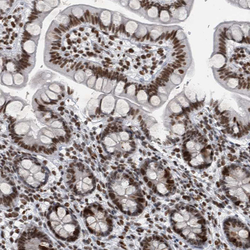
- Immunohistochemical staining of human gastrointestinal shows strong nuclear positivity in glandular cells.
- Sample type
- HUMAN

- Submitted by
- Atlas Antibodies (provider)
- Main image
- Experimental details
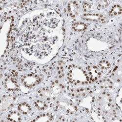
- Immunohistochemical staining of human kidney shows moderate nuclear positivity in cells in glomeruli and tubules.
- Sample type
- HUMAN

- Submitted by
- Atlas Antibodies (provider)
- Main image

- Experimental details
- Immunohistochemical staining of human pancreas shows weak to moderate nuclear positivity in islets of Langerhans and exocrine glandular cells.
- Sample type
- HUMAN
