 Explore
 Validate
 Learn
Learn Western blot
Antibody data
- Antibody Data
- Antigen structure
- References [1]
- Comments [0]
- Validations
- Western blot [2]
Submit
Validation data
Reference
Comment
Report error
- Product number
- MAB8279 - Provider product page

- Provider
- Novus Biologicals
- Product name
- Mouse Monoclonal alpha-Actinin 1 Antibody
- Antibody type
- Monoclonal
- Description
- Protein A or G purified from hybridoma culture supernatant. Detects human alpha-Actinin 1 in ELISA, and human, mouse and rat alpha-Actinin 1 in Western Blot.
- Reactivity
- Human, Mouse, Rat
- Host
- Mouse
- Conjugate
- Unconjugated
- Isotype
- IgG
- Vial size
- 100 ug
- Concentration
- LYOPH
- Storage
- Use a manual defrost freezer and avoid repeated freeze-thaw cycles. 12 months from date of receipt, -20 to -70 degreesC as supplied. 1 month, 2 to 8 degreesC under sterile conditions after reconstitution. 6 months, -20 to -70 degreesC under sterile conditions after reconstitution.
Submitted references Indirect Chronic Effects of an Oleuropein-Rich Olive Leaf Extract on Sucrase-Isomaltase In Vitro and In Vivo.
Pyner A, Chan SY, Tumova S, Kerimi A, Williamson G
Nutrients 2019 Jul 1;11(7)
Nutrients 2019 Jul 1;11(7)
No comments: Submit comment
Supportive validation
- Submitted by
- Novus Biologicals (provider)
- Main image
- Experimental details
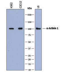
- Detection of Human, Mouse, and Rat alpha-Actinin 1 by Western Blot. Western blot shows lysates of K562 human chronic myelogenous leukemia cell line, C2C12 mouse myoblast cell line, and L6 rat myoblast cell line. PVDF membrane was probed with 0.2 µg/mL of Mouse Anti-Human/Mouse/Rat alpha-Actinin 1 Monoclonal Antibody (Catalog # MAB8279) followed by HRP-conjugated Anti-Mouse IgG Secondary Antibody (Catalog # HAF018). A specific band was detected for alpha-Actinin 1 at approximately 100-105 kDa (as indicated). This experiment was conducted under reducing conditions and using Immunoblot Buffer Group 1.

- Submitted by
- Novus Biologicals (provider)
- Main image
- Experimental details
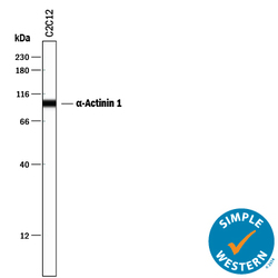
- Detection of Mouse alpha-Actinin 1 by Simple WesternTM. Simple Western lane view shows lysates of C2C12 mouse myoblast cell line, loaded at 0.5 mg/mL. A specific band was detected for alpha-Actinin 1 at approximately 98 kDa (as indicated) using 2 µg/mL of Mouse Anti-Human/Mouse/Rat alpha-Actinin 1 Monoclonal Antibody (Catalog # MAB8279). This experiment was conducted under reducing conditions and using the 12-230 kDa separation system.
