 Explore
 Validate
 Learn
Learn Immunohistochemistry
Antibody data
- Antibody Data
- Antigen structure
- References [20]
- Comments [0]
- Validations
- Immunohistochemistry [1]
- Other assay [9]
Submit
Validation data
Reference
Comment
Report error
- Product number
- PA1-26938 - Provider product page

- Provider
- Invitrogen Antibodies
- Product name
- Insulin Polyclonal Antibody
- Antibody type
- Polyclonal
- Antigen
- Other
- Description
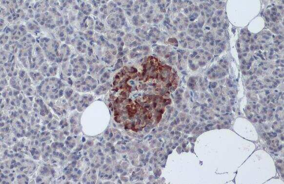
- This antibody stains beta(b) cells of the Langerhan's islets in human pancreas. IHC Note: prolonged fixation in buffered formalin can destroy the epitope.
- Reactivity
- Human, Mouse
- Host
- Guinea Pig
- Isotype
- IgG
- Vial size
- 500 µL
- Concentration
- 0.2 mg/mL
- Storage
- 4° C, do not freeze
Submitted references Evidence of islet CADM1-mediated immune cell interactions during human type 1 diabetes.
The IGFBP3/TMEM219 pathway regulates beta cell homeostasis.
Integrated Array Tomography for 3D Correlative Light and Electron Microscopy.
Insm1, Neurod1, and Pax6 promote murine pancreatic endocrine cell development through overlapping yet distinct RNA transcription and splicing programs.
A developmental lineage-based gene co-expression network for mouse pancreatic β-cells reveals a role for Zfp800 in pancreas development.
Maternal adaptations of pancreatic islets and glucose metabolism after lactation.
Vertical sleeve gastrectomy triggers fast β-cell recovery upon overt diabetes.
Insulinoma-derived pseudo-islets for diabetes research.
Scrt1, a transcriptional regulator of β-cell proliferation identified by differential chromatin accessibility during islet maturation.
A monoclonal antibody with broad specificity for the ligands of insulin B:9-23 reactive T cells prevents spontaneous type 1 diabetes in mice.
Oligomeric collagen as an encapsulation material for islet/β-cell replacement: effect of islet source, dose, implant site, and administration format.
Macrophage IFN-I signaling promotes autoreactive T cell infiltration into islets in type 1 diabetes model.
In situ type I oligomeric collagen macroencapsulation promotes islet longevity and function in vitro and in vivo.
Nrf2 prevents Notch-induced insulin resistance and tumorigenesis in mice.
GRP94 Is an Essential Regulator of Pancreatic β-Cell Development, Mass, and Function in Male Mice.
Long noncoding RNAs are dynamically regulated during β-cell mass expansion in mouse pregnancy and control β-cell proliferation in vitro.
β-arrestin-2 is an essential regulator of pancreatic β-cell function under physiological and pathophysiological conditions.
Hypothyroidism Impairs Human Stem Cell-Derived Pancreatic Progenitor Cell Maturation in Mice.
Characterization of age-associated alterations of islet function and structure in diabetic mutant cryptochrome 1 transgenic mice.
Mitochondrial dysfunction and increased reactive oxygen species impair insulin secretion in sphingomyelin synthase 1-null mice.
Sona C, Yeh YT, Patsalos A, Halasz L, Yan X, Kononenko NL, Nagy L, Poy MN
JCI insight 2022 Mar 22;7(6)
JCI insight 2022 Mar 22;7(6)
The IGFBP3/TMEM219 pathway regulates beta cell homeostasis.
D'Addio F, Maestroni A, Assi E, Ben Nasr M, Amabile G, Usuelli V, Loretelli C, Bertuzzi F, Antonioli B, Cardarelli F, El Essawy B, Solini A, Gerling IC, Bianchi C, Becchi G, Mazzucchelli S, Corradi D, Fadini GP, Foschi D, Markmann JF, Orsi E, Škrha J Jr, Camboni MG, Abdi R, James Shapiro AM, Folli F, Ludvigsson J, Del Prato S, Zuccotti G, Fiorina P
Nature communications 2022 Feb 3;13(1):684
Nature communications 2022 Feb 3;13(1):684
Integrated Array Tomography for 3D Correlative Light and Electron Microscopy.
Lane R, Wolters AHG, Giepmans BNG, Hoogenboom JP
Frontiers in molecular biosciences 2021;8:822232
Frontiers in molecular biosciences 2021;8:822232
Insm1, Neurod1, and Pax6 promote murine pancreatic endocrine cell development through overlapping yet distinct RNA transcription and splicing programs.
Dudek KD, Osipovich AB, Cartailler JP, Gu G, Magnuson MA
G3 (Bethesda, Md.) 2021 Oct 19;11(11)
G3 (Bethesda, Md.) 2021 Oct 19;11(11)
A developmental lineage-based gene co-expression network for mouse pancreatic β-cells reveals a role for Zfp800 in pancreas development.
Osipovich AB, Dudek KD, Greenfest-Allen E, Cartailler JP, Manduchi E, Potter Case L, Choi E, Chapman AG, Clayton HW, Gu G, Stoeckert CJ Jr, Magnuson MA
Development (Cambridge, England) 2021 Mar 21;148(6)
Development (Cambridge, England) 2021 Mar 21;148(6)
Maternal adaptations of pancreatic islets and glucose metabolism after lactation.
Canul-Medina G, Riverón-Negrete L, Pastén-Hidalgo K, Morales-Castillo P, García-Vázquez F, Fernandez-Mejia C
The Journal of endocrinology 2021 Jan;248(1):1-15
The Journal of endocrinology 2021 Jan;248(1):1-15
Vertical sleeve gastrectomy triggers fast β-cell recovery upon overt diabetes.
Oppenländer L, Palit S, Stemmer K, Greisle T, Sterr M, Salinno C, Bastidas-Ponce A, Feuchtinger A, Böttcher A, Ansarullah, Theis FJ, Lickert H
Molecular metabolism 2021 Dec;54:101330
Molecular metabolism 2021 Dec;54:101330
Insulinoma-derived pseudo-islets for diabetes research.
Hart NJ, Weber C, Price N, Banuelos A, Schultz M, Huey B, Harnois E, Gibson C, Steyn LV, Papas KK, Lynch RM
American journal of physiology. Cell physiology 2021 Aug 1;321(2):C247-C256
American journal of physiology. Cell physiology 2021 Aug 1;321(2):C247-C256
Scrt1, a transcriptional regulator of β-cell proliferation identified by differential chromatin accessibility during islet maturation.
Sobel J, Guay C, Elhanani O, Rodriguez-Trejo A, Stoll L, Menoud V, Jacovetti C, Walker MD, Regazzi R
Scientific reports 2021 Apr 22;11(1):8800
Scientific reports 2021 Apr 22;11(1):8800
A monoclonal antibody with broad specificity for the ligands of insulin B:9-23 reactive T cells prevents spontaneous type 1 diabetes in mice.
Cepeda JR, Sekhar NS, Han J, Xiong W, Zhang N, Yu L, Dai S, Davidson HW, Kappler JW, An Z, Zhang L
mAbs 2020 Jan-Dec;12(1):1836714
mAbs 2020 Jan-Dec;12(1):1836714
Oligomeric collagen as an encapsulation material for islet/β-cell replacement: effect of islet source, dose, implant site, and administration format.
Stephens CH, Morrison RA, McLaughlin M, Orr K, Tersey SA, Scott-Moncrieff JC, Mirmira RG, Considine RV, Voytik-Harbin S
American journal of physiology. Endocrinology and metabolism 2020 Aug 1;319(2):E388-E400
American journal of physiology. Endocrinology and metabolism 2020 Aug 1;319(2):E388-E400
Macrophage IFN-I signaling promotes autoreactive T cell infiltration into islets in type 1 diabetes model.
Marro BS, Legrain S, Ware BC, Oldstone MB
JCI insight 2019 Jan 24;4(2)
JCI insight 2019 Jan 24;4(2)
In situ type I oligomeric collagen macroencapsulation promotes islet longevity and function in vitro and in vivo.
Stephens CH, Orr KS, Acton AJ, Tersey SA, Mirmira RG, Considine RV, Voytik-Harbin SL
American journal of physiology. Endocrinology and metabolism 2018 Oct 1;315(4):E650-E661
American journal of physiology. Endocrinology and metabolism 2018 Oct 1;315(4):E650-E661
Nrf2 prevents Notch-induced insulin resistance and tumorigenesis in mice.
Chartoumpekis DV, Yagishita Y, Fazzari M, Palliyaguru DL, Rao UN, Zaravinos A, Khoo NK, Schopfer FJ, Weiss KR, Michalopoulos GK, Sipula I, O'Doherty RM, Kensler TW, Wakabayashi N
JCI insight 2018 Mar 8;3(5)
JCI insight 2018 Mar 8;3(5)
GRP94 Is an Essential Regulator of Pancreatic β-Cell Development, Mass, and Function in Male Mice.
Kim DS, Song L, Wang J, Wu H, Gu G, Sugi Y, Li Z, Wang H
Endocrinology 2018 Feb 1;159(2):1062-1073
Endocrinology 2018 Feb 1;159(2):1062-1073
Long noncoding RNAs are dynamically regulated during β-cell mass expansion in mouse pregnancy and control β-cell proliferation in vitro.
Sisino G, Zhou AX, Dahr N, Sabirsh A, Soundarapandian MM, Perera R, Larsson-Lekholm E, Magnone MC, Althage M, Tyrberg B
PloS one 2017;12(8):e0182371
PloS one 2017;12(8):e0182371
β-arrestin-2 is an essential regulator of pancreatic β-cell function under physiological and pathophysiological conditions.
Zhu L, Almaça J, Dadi PK, Hong H, Sakamoto W, Rossi M, Lee RJ, Vierra NC, Lu H, Cui Y, McMillin SM, Perry NA, Gurevich VV, Lee A, Kuo B, Leapman RD, Matschinsky FM, Doliba NM, Urs NM, Caron MG, Jacobson DA, Caicedo A, Wess J
Nature communications 2017 Feb 1;8:14295
Nature communications 2017 Feb 1;8:14295
Hypothyroidism Impairs Human Stem Cell-Derived Pancreatic Progenitor Cell Maturation in Mice.
Bruin JE, Saber N, O'Dwyer S, Fox JK, Mojibian M, Arora P, Rezania A, Kieffer TJ
Diabetes 2016 May;65(5):1297-309
Diabetes 2016 May;65(5):1297-309
Characterization of age-associated alterations of islet function and structure in diabetic mutant cryptochrome 1 transgenic mice.
Okano S, Hayasaka K, Igarashi M, Togashi Y, Nakajima O
Journal of diabetes investigation 2013 Sep 13;4(5):428-35
Journal of diabetes investigation 2013 Sep 13;4(5):428-35
Mitochondrial dysfunction and increased reactive oxygen species impair insulin secretion in sphingomyelin synthase 1-null mice.
Yano M, Watanabe K, Yamamoto T, Ikeda K, Senokuchi T, Lu M, Kadomatsu T, Tsukano H, Ikawa M, Okabe M, Yamaoka S, Okazaki T, Umehara H, Gotoh T, Song WJ, Node K, Taguchi R, Yamagata K, Oike Y
The Journal of biological chemistry 2011 Feb 4;286(5):3992-4002
The Journal of biological chemistry 2011 Feb 4;286(5):3992-4002
No comments: Submit comment
Supportive validation
- Submitted by
- Invitrogen Antibodies (provider)
- Main image

- Experimental details
- Immunohistochemistry (Paraffin) analysis of Insulin was performed in paraffin-embedded human pancreas tissue using Insulin Polyclonal Antibody (Product # PA1-26938) at a dilution of 1:50. Antigen Retrieval: Citrate buffer, pH 6.0, 15 min.
Supportive validation
- Submitted by
- Invitrogen Antibodies (provider)
- Main image

- Experimental details
- NULL
- Submitted by
- Invitrogen Antibodies (provider)
- Main image

- Experimental details
- NULL

- Submitted by
- Invitrogen Antibodies (provider)
- Main image

- Experimental details
- NULL

- Submitted by
- Invitrogen Antibodies (provider)
- Main image

- Experimental details
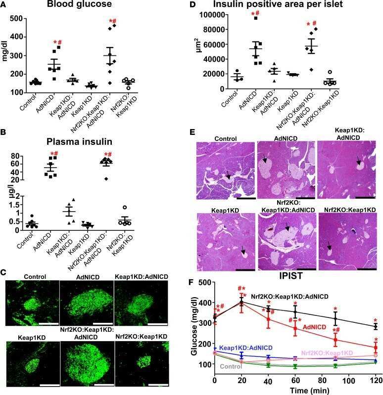
- Figure 2 Enhancer activity assessment and Scrt1 function. ( a ) Trn5 integrations on Mafb, Syt4, Neurod1 and two Pax6 accessible sites. ( b ) Scheme of pGL3 vector used for the luciferase assay. ( c ) Luciferase activity was measured in INS 832/13 cells transfected with an empty pGL3 vector (Ctrl) or a pGL3 vector containing an enhancer region for the indicated gene ( Mafb, NeuroD1 , Pax6 and Syt4 ). Results are expressed as fold change versus control. ( d ) Gene expression in P10 and adult rat islets were measured by qPCR and normalized to the housekeeping gene Hprt1 . Syt4 gene expression is available in Fig. 3 . * p < 0.05, ** p < 0.01 by Student''s t-test or by one-way Anova, Dunnett''s post-hoc test. ( e - f ) Scrt1 expression was measured by qPCR and normalized to Hprt1 housekeeping gene levels. ( f - j ) Dispersed adult rat islet cells were transfected with a control siRNA (siCtl) or siRNAs directed against Scrt1 (siScrt1). Experiments were performed 48 h post-transfection. Insulin release in response to 2 or 20 mM glucose ( g,h ) insulin content were determined by ELISA n = 3. ( i ) Apoptosis of insulin-positive cells was assessed using Tunel assay in basal (NT) condition or in response to a mix (cyt mix) of pro-inflammatory cytokines (IL-1beta, TNF-alpha and IFN-gamma) n = 5. ( j ) The fraction of proliferative insulin-positive cells was determined by BrdU incorporation in basal (NT) or stimulated (prolactin, PRL) conditions, n = 6. * p < 0.05, ** p < 0.01 by Student

- Submitted by
- Invitrogen Antibodies (provider)
- Main image
- Experimental details
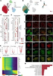
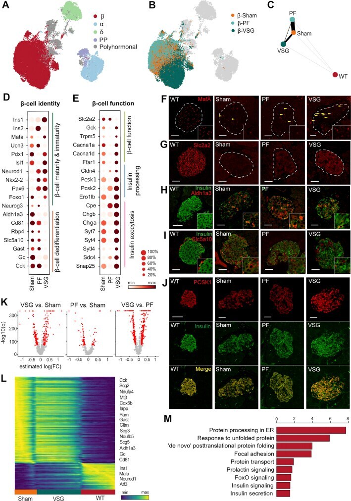
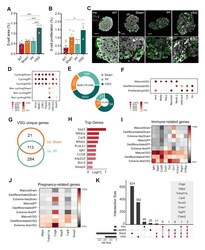
- Figure 3 VSG distinctly improves the identity and function of beta-cells . A,B , UMAP plots of 35,941 endocrine cells from Sham, VSG and PF. Colours in (a) indicate the five main endocrine cell clusters and in (b) distinguish beta-cells specifically according to the three groups. C , Abstracted graph of cluster connectivity assessing transcriptomic similarity between VSG-, PF-, Sham- and WT-derived beta-cells, inferred using PAGA. PAGA is based on a statistical method that measures relatedness between single-cell clusters, with edge weights indicating link significance. D,E , Gene expression for key genes involved in beta-cell identity and function changed upon VSG. (d) Dot plot showing that VSG-derived beta-cells upregulated essential maturity markers and downregulated dedifferentiation markers. (e) Dot plot showing genes critical for beta-cell function and metabolism, insulin processing and exocytosis. Color intensity shows normalized mean expression in a group; dot size indicates the percentage of cells that express the corresponding gene. Expression is scaled per gene. F-J , Immunohistochemical analysis of MafA, Slc2a2, Insulin, Aldh1a3, Slc5a10 and Pcsk1 in beta-cells of WT, Sham, PF and VSG mice at study end. Scale is 50 mum K , Volcano plots indicating differences in the number of genes that were differentially expressed when comparing beta-cells from Sham and VSG versus beta-cells from Sham and PF, obtained from differential analysis testing using limma-trend. Red dot
- Submitted by
- Invitrogen Antibodies (provider)
- Main image
- Experimental details
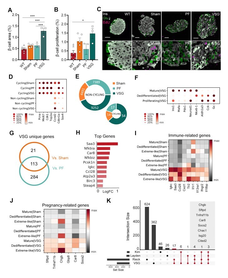
- Figure 6 VSG rapidly expands beta-cell mass . A , Pancreas morphometry analysis reveals that VSG doubled the relative beta-cell area (WT, n = 6; Sham, n = 5, PF, n = 3; VSG, n = 3; WT versus VSG (p < 0.0001); Sham versus VSG (p < 0.0001), PF vs VSG (p = 0.0003)). B , Quantification of beta-cell proliferation at study end (WT, n = 6; Sham, n = 5, PF, n = 3; VSG, n = 3; WT versus VSG (p = 0.019), VSG versus Sham (p > 0.999), VSG versus PF (p = 0.156)). All data are presented as mean +- s.e.m and were analyzed by one-way ANOVA with Bonferroni's post-hoc test. C , Representative images of immuno-stainings against insulin, glucagon and EdU. Scale is 50 mum D , Dot plot showing expression of cell cycle-related genes in cycling and non-cycling beta-cells per group. Color intensity shows normalized mean expression in a cell cluster. Expression is scaled per gene. E , Doughnut plots indicating fraction of proliferating beta-cells per group (VSG-54/9252, PF-17/7389, Sham-14/4491). F , Dot plot showing expression of beta-cell identity genes in mature, dedifferentiated and proliferating beta-cells of VSG, implying the dedifferentiated signature of proliferating beta-cells. Expression is scaled per gene. G , Venn diagram showing uniquely upregulated beta-cell genes in VSG versus control groups. H , Top 10 unique VSG genes (estimated log (FC) > 0.25, B > 150). Log (FC) plotted for VSG versus Sham. I-J , Matrix plots showing expression of (i) immune-related and (j) pregnancy-related genes i
- Submitted by
- Invitrogen Antibodies (provider)
- Main image
- Experimental details
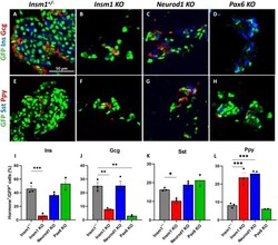
- Figure 2 Impaired differentiation of pancreatic endocrine cells in Insm1 , Neurod1 , and Pax6 KO embryos. (A-D) Immunofluorescence labeling of pancreata from E18.5 Insm1 GFP -expressing embryos using antibodies against GFP (green) that marks pre-endocrine cells, and pancreatic hormones insulin (Ins, blue), and glucagon (Gcg, red). Compared with Insm1 +/- mice (A), mice lacking Insm1 (B), Neurod1 (C), and Pax6 (D) exhibit a decrease in total number of endocrine cells, altered endocrine cell morphology and numbers of hormone expressing cells. (E-H) Immunofluorescence labeling of pancreata from E18.5 embryos using antibodies against GFP (green), pancreatic polypeptide (Ppy, red), and somatostatin (Sst, blue) shows altered numbers of hormone expressing cells in Insm1 (F), Neurod1 (G), and Pax6 (H) KO mice. (I-L) Quantification of a percentage of hormone-positive cells among GFP-positive endocrine cells demonstrates defects in differentiation of cells positive for hormones: insulin (Ins) (I), glucagon (Gcg) (J), somatostatin (Sst) (K), and pancreatic polypeptide (Ppy) (L) in Insm1 , Neurod1 , and Pax6 KO embryonic pancreata in comparison with Insm1 +/- . Error bars indicate SEM ( n = 3); P -values were determined by one-way ANOVA test. Asterisks indicate P -values of *

- Submitted by
- Invitrogen Antibodies (provider)
- Main image
- Experimental details
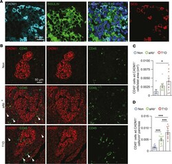
- Increased number of CADM1 + CD45 + cells within the pancreatic islet during T1D. ( A ) Immunostaining of paraffin-embedded pancreata from individuals in the Non group for CADM1 (cyan), insulin (green), and GCG (red). Scale bar: 20 mum. ( B ) Immunostaining of paraffin-embedded pancreata from individuals in the Non, aAb + , and T1D groups for CADM1 (red) and CD45 (green). Scale bar: 50 mum. ( C ) Quantification of the number of CADM1 + CD45 + cells within the islet periphery ( n = 5 per group). ( D ) Quantification of the number of CD45 + adjacent to CADM1 + cells per area pancreas ( n = 5 per group). One-way ANOVA was performed using GraphPad Prism, version 7, software for comparisons of 3 groups. Post hoc statistical analyses were performed using the Tukey multiple comparisons test. Results are presented as mean +- SEM. * P < 0.05; *** P < 0.001. ad., adjacent.

- Submitted by
- Invitrogen Antibodies (provider)
- Main image
- Experimental details
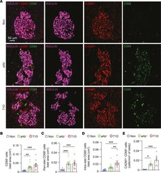
- Increased number of CD68 + cells adjacent to CADM1 + insulin + cells during T1D. ( A ) Immunostaining of paraffin-embedded pancreata from individuals in the Non, aAb + , and T1D groups for CADM1 (red), CD68 (green), and insulin (magenta). Scale bar: 50 mum. ( B ) Quantification of the number of CD68 + cells within the islet boundary ( n = 5 per group). ( C ) Quantification of the number of CD68 + cells at the islet periphery per islet area ( n = 5 per group). ( D ) Quantification of the number of CD68 + cells within the islet boundary ( n = 5 per group). ( E ) Quantification of the number of CADM1 + CD68 + cells within the islet boundary ( n = 5 per group). One-way ANOVA was performed using GraphPad Prism, version 7, software for comparisons of 3 groups. Post hoc statistical analyses were performed using Tukey's multiple comparisons test. Results are presented as mean +- SEM. * P < 0.05; ** P < 0.01; *** P < 0.001.
