 Explore
 Validate
 Learn
Learn Western blot
 Immunocytochemistry
Antibody data
- Antibody Data
- Antigen structure
- References [3]
- Comments [0]
- Validations
- Immunocytochemistry [3]
- Chromatin Immunoprecipitation [1]
- Other assay [5]
Submit
Validation data
Reference
Comment
Report error
- Product number
- PA5-17876 - Provider product page

- Provider
- Invitrogen Antibodies
- Product name
- Phospho-STAT3 (Ser727) Polyclonal Antibody
- Antibody type
- Polyclonal
- Antigen
- Synthetic peptide
- Description
- It is not recommended to aliquot this antibody.
- Reactivity
- Human, Mouse, Rat
- Host
- Rabbit
- Isotype
- IgG
- Vial size
- 100 µL
- Concentration
- 130 µg/mL
- Storage
- -20°C
Submitted references Krüppel-like factor 7 attenuates hippocampal neuronal injury after traumatic brain injury.
STAT3-mediated allelic imbalance of novel genetic variant Rs1047643 and B-cell-specific super-enhancer in association with systemic lupus erythematosus.
Respiratory syncytial virus reduces STAT3 phosphorylation in human memory CD8 T cells stimulated with IL-21.
Li WY, Fu XM, Wang ZD, Li ZG, Ma D, Sun P, Liu GB, Zhu XF, Wang Y
Neural regeneration research 2022 Mar;17(3):661-672
Neural regeneration research 2022 Mar;17(3):661-672
STAT3-mediated allelic imbalance of novel genetic variant Rs1047643 and B-cell-specific super-enhancer in association with systemic lupus erythematosus.
Zhang Y, Day K, Absher DM
eLife 2022 Feb 21;11
eLife 2022 Feb 21;11
Respiratory syncytial virus reduces STAT3 phosphorylation in human memory CD8 T cells stimulated with IL-21.
Antunes KH, Becker A, Franceschina C, de Freitas DDN, Lape I, da Cunha MD, Leitão L, Rigo MM, Pinto LA, Stein RT, de Souza APD
Scientific reports 2019 Nov 28;9(1):17766
Scientific reports 2019 Nov 28;9(1):17766
No comments: Submit comment
Supportive validation
- Submitted by
- Invitrogen Antibodies (provider)
- Main image

- Experimental details
- Immunofluorescence analysis of Phospho-STAT3 (Ser727) was performed using 90% confluent log phase NIH/3T3 cells treated with IL6 (100 ng/mL, 30 mins). The cells were fixed with 4% paraformaldehyde for 10 minutes, permeabilized with 0.1% Triton™ X-100 for 15 minutes, and blocked with 1% BSA for 1 hour at room temperature. The cells were labeled with Phospho-STAT3 (Ser727) Polyclonal Antibody (Product # PA5-17876) at 1:100 in 0.1% BSA, incubated at 4 degree Celsius overnight and then labeled with Goat anti-Rabbit IgG (H+L) Superclonal™ Secondary Antibody, Alexa Fluor® 488 conjugate (Product # A27034) at a dilution of 1:2000 for 45 minutes at room temperature (Panel a: green). Nuclei (Panel b: blue) were stained with SlowFade® Gold Antifade Mountant with DAPI (Product # S36938). F-actin (Panel c: red) was stained with Rhodamine Phalloidin (Product # R415, 1:300). Panel d represents the merged image showing nuclear localization. Panel e represents untreated cells. Panel f represents control cells with no primary antibody to assess background. The images were captured at 60X magnification.

- Submitted by
- Invitrogen Antibodies (provider)
- Main image

- Experimental details
- Immunofluorescent analysis of Phospho-STAT3 pSer727 in HeLa cells, IL-6 treated, using a Phospho-STAT3 pSer727 polyclonal antibody (Product # PA5-17876) (green) and a Pan-Keratin monoclonal antibody (red).

- Submitted by
- Invitrogen Antibodies (provider)
- Main image

- Experimental details
- Immunofluorescent analysis of Phospho-STAT3 pSer727 in HeLa cells using a Phospho-STAT3 pSer727 polyclonal antibody (Product # PA5-17876) (green) and a Pan-Keratin monoclonal antibody (red).

Supportive validation
- Submitted by
- Invitrogen Antibodies (provider)
- Main image

- Experimental details
- Enrichment of endogenous Phospho-STAT3 (Ser727) protein at specific gene loci using Anti-Phospho-STAT3 (Ser727) Antibody: Chromatin Immunoprecipitation (ChIP) was performed using Anti-Phospho-STAT3 (Ser727) Rabbit Polyclonal Antibody (Product # PA5-17876, 8 ul) on sheared chromatin from 2 million HeLa cells treated with IL-6 (100 ng/ml for 30 min) using the MAGnify ChIP System (Product # 49-2024). Normal Rabbit IgG was used as a negative IP control. The purified DNA was analyzed by qPCR with PCR primer pairs over IRF1, FOS, SOCS3 (active) and SAT2 satellite repeats and SAT alpha (inactive). Data is presented as fold enrichment of the antibody signal versus the negative control IgG using the comparative CT method.

Supportive validation
- Submitted by
- Invitrogen Antibodies (provider)
- Main image
- Experimental details
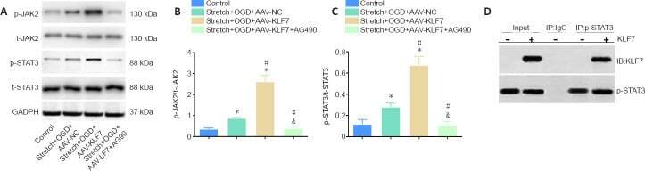
- Figure 4 Involvement of the JAK2/STAT3 signaling pathway in the neuroprotective effects of KLF7. (A) Representative western blot of p-JAK2, t-JAK2, p-STAT3, and t-STAT3 expression in cultured HT22 cells at 1 day after stretch and OGD treatment. (B, C) Quantitative results of the relative optical densities of p-JAK2/t-JAK2 and p-STAT3/t-STAT3. (D) Co-immunoprecipitation analysis of the interactions between KLF7 and p-STAT3 in stretch- and OGD-damaged HT22 cells at 1 day, revealing a physical interaction between KLF7 and p-STAT3. Data are expressed as the mean +- SD. Each experiment was repeated three times. * P < 0.05, vs . control group; # P < 0.05, vs . stretch + OGD + AAV-NC group; & P < 0.05, vs . stretch + OGD + AAV-KLF7 group (one-way analysis of variance followed by Tukey's post hoc test). AAV: Adeno-associated virus; GAPDH: glyceraldehyde-3-phosphate dehydrogenase; IB: immunoblotting; IP: immunoprecipitation; KLF7: Kruppel-like factor 7; NC: negative control; OGD: oxygen-glucose deprivation; p-JAK2: phospho-Janus kinase 2; t-JAK2: total-Janus kinase 2; p-STAT3: phospho-signal transducer and activator of transcription 3; t-STAT3: total-signal transducer and activator of transcription 3.
- Submitted by
- Invitrogen Antibodies (provider)
- Main image
- Experimental details
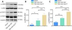
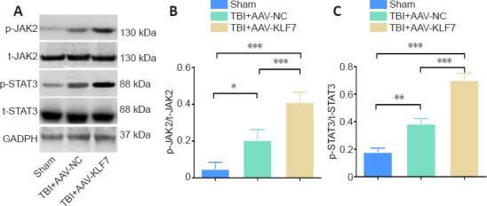
- Figure 9 KLF7 activates the JAK2/STAT3 pathway at 3 days after TBI. (A) Representative western blots of p-JAK2, t-JAK2, p-STAT3, and t-STAT3 expression in ipsilateral hippocampal tissue. (B, C) Quantitative results of the p-JAK2/t-JAK2 and p-STAT3/t-STAT3 ratios. Data are expressed as the mean +- SD ( n = 3). * P < 0.05, ** P < 0.01, *** P < 0.001 (one-way analysis of variance followed by Tukey's post hoc test). AAV: Adeno-associated virus; GAPDH: glyceraldehyde-3-phosphate dehydrogenase; KLF7: Kruppel-like factor 7; NC: negative control; p-JAK2: phospho-Janus kinase 22; p-STAT3: phospho-signal transducer and activator of transcription 3; t-JAK2: total-Janus kinase; t-STAT3: total-signal transducer and activator of transcription 3; TBI: traumatic brain injury.
- Submitted by
- Invitrogen Antibodies (provider)
- Main image

- Experimental details
- Figure 2 RSV inhibits pSTAT3 induced by IL-21 in purified human memory CD8 T cells. Human memory CD8 T cells were isolated from PBMCs, incubated with RSV (5 x 10² PFU/ml) for 1 h and treated with IL-21 (25 ng/ml). After 30 min, the cells were harvested, fixed and stained for immunofluorescence analysis. ( A ) Fluorescence images of cell nuclei stained for Hoechst (blue) and pSTAT3 Ser727 (red). ( B ) Quantification of STAT3 phosphorylation on Ser727in purified human CD8 T cells.

- Submitted by
- Invitrogen Antibodies (provider)
- Main image

- Experimental details
- Figure 3 RSV inhibits pSTAT3 induced by IL-21 in live purified human memory CD8 T cells. Human memory CD8 T cells were isolated from PBMCs, incubated with RSV (5 x 10² PFU/ml) for 1 h and treated with IL-21 (25 ng/ml). After 30 min, the cells were harvested and stained for flow cytometry analysis. ( A ) Gate strategy and representative plots of flow cytometry analysis of pSTAT3 in memory CD8 T cells. ( B ) MFI (mean of fluorescence intensity) of pSTAT3 in memory CD8 T cells. Data are expressed as the mean +- SEM. Statistical significance was determined using one-way ANOVA followed by Tukey's multiple comparison test. * p < 0.05, ** p < 0.01, *** p < 0.001.

- Submitted by
- Invitrogen Antibodies (provider)
- Main image

- Experimental details
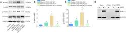
- Figure 6 Pulmonary cells infected with RSV inhibit STAT3 phosphorylation and cytotoxic activity in IL-21-treated human CD8 T cells. Human pulmonary cells (MRC5) were infected with RSV (1 x 10 4 PFU/ml) for 1 h. Afterwards, the medium was replenished, and human CD8 T cells isolated from PBMCs were placed in coculture with the infected MRC5 cells and then stimulated with IL-21 (25 ng/ml) for 30 min. ( A ) Cropped blot bands of total phosphorylated STAT3 protein (on serine 727) in memory CD8 T cells analyzed by Western blot. beta-Actin was used to normalize protein quantification. ( B ) pSTAT3 protein quantification from Western blot analysis. Data are shown as percentages over untreated/uninfected controls. Full-length blots are shown in Supplementary Fig. 4 . ( C ) Human pulmonary MRC-5 cells (1 x 10 5 cells) were infected with RSV and cocultured in 96-well plates with human memory CD8 T cells (8 x 10 4 cells) stimulated with IL-21 (25 ng/ml) in RPMI with 2% FBS. Human CD8 T cell cytotoxicity against MRC-5 cells was assessed by lactate dehydrogenase detection in the coculture supernatant. Data are expressed as the mean +- SEM. Statistical significance was determined using one-way ANOVA followed by Tukey's multiple comparison test. * p < 0.05, ** p < 0.01, *** p < 0.001.
